. 2012 May;86(9):5134-50.
doi: 10.1128/JVI.07137-11. Epub 2012 Feb 29.

In vitro epsilon RNA-dependent protein priming activity of human hepatitis B virus polymerase

Affiliations

In vitro epsilon RNA-dependent protein priming activity of human hepatitis B virus polymerase

Scott A Jones et al. J Virol. 2012 May.

Erratum in

  • J Virol. 2013 Nov;87(22):12504

Abstract

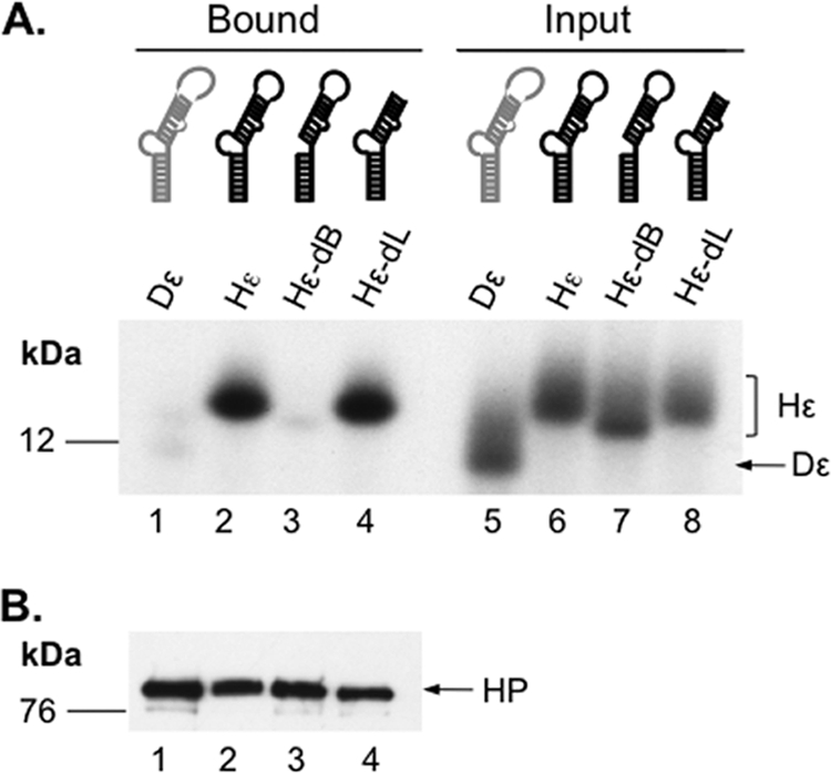
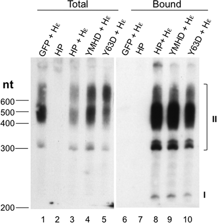
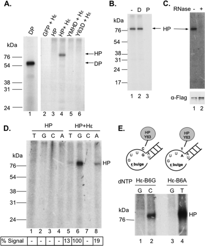
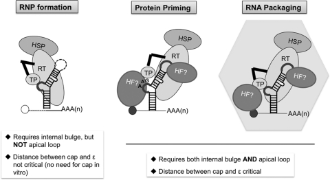
Hepatitis B virus (HBV) replicates its DNA genome through reverse transcription of a pregenomic RNA (pgRNA) by using a multifunctional polymerase (HP). A critical function of HP is its specific recognition of a viral RNA signal termed ε (Hε) located on pgRNA, which is required for specific packaging of pgRNA into viral nucleocapsids and initiation of viral reverse transcription. HP initiates reverse transcription by using itself as a protein primer (protein priming) and Hε as the obligatory template. We have purified HP from human cells that retained Hε binding activity in vitro. Furthermore, HP purified as a complex with Hε, but not HP alone, displayed in vitro protein priming activity. While the HP-Hε interaction in vitro and in vivo required the Hε internal bulge, but not its apical loop, and was not significantly affected by the cap-Hε distance, protein priming required both the Hε apical loop and internal bulge, as well as a short distance between the cap and Hε, mirroring the requirements for RNA packaging. These studies have thus established new HBV protein priming and RNA binding assays that should greatly facilitate the dissection of the requirements and molecular mechanisms of HP-Hε interactions, RNA packaging, and protein priming.


Figures

Fig 1
Fig 1
Purification of HBV polymerase (HP) in association with host factors. The 3×FLAG-tagged HP was purified with the M2 (anti-FLAG [α-FLAG])-bound protein A/G beads from transiently transfected HEK293T cells (A, lane 3, and B, lane 2). HP + Hε represents purification of HP from cells that were coexpressing the Hε RNA (A, lane 4). Control purification in parallel was performed with GFP-transfected cells (A, lane 2; B, lane 1). (A) Purified HP and bound cellular proteins were resolved on an SDS–10% polyacrylamide gel and visualized by silver staining. (B) The identity of HP and selected host proteins was verified by Western blotting using the indicated antibodies (α-). The positions of the protein molecular mass markers (in kDa) are indicated (lane M), as are the various host proteins and the antibody heavy and light chains in panel A. The symbol * in panel B indicates the antibody heavy chain that cross-reacted weakly with anti-Hsp60.
Fig 2
Fig 2
Detection of HP-Hε interaction in vitro. Immunoaffinity-purified HP was incubated with 32P-labeled, in vitro-transcribed WT or mutant Hε or Dε RNA. After removing unbound RNA, the bound RNA and HP were resolved on an SDS–15% polyacrylamide gel. (A) The bottom portion of the gel, which contained the 32P-labeled ε RNA, was dried and analyzed by autoradiography (lanes 1 to 4). Input representing 1% of the amount of the indicated ε RNA added to each binding reaction mixture was also analyzed in parallel (lanes 5 to 8). (B) The top portion of the gel containing HP was analyzed by Western blotting for HP in the binding reactions using the M2 anti-FLAG antibody. The Hε and Dε RNAs and HP are indicated, as are the positions of the protein molecular mass markers (in kDa).
Fig 3
Fig 3
Detection of HP-Hε interaction in vivo. Total RNA was extracted from HEK293T cells transfected with the indicated construct(s) (lanes 1 to 5). HP-bound RNA was extracted from immunoaffinity-purified HP (lanes 6 to 10). The purified RNAs were resolved on an 8 M urea–6% polyacrylamide gel, electrotransferred to membrane, and probed with a 32P-labeled riboprobe specific for the Hε RNA sequence. The capped, polyadenylated Hε species are indicated (I and II): I, ca. 200 nucleotides long, representing polyadenylation at the weak native HBV poly(A) site that is ca. 100 nucleotides downstream of the 5′ cap; II, from 300 to 600 nucleotides long, representing polyadenylation at the strong bovine growth hormone (BGH) poly(A) site ca. 220 nucleotides downstream from the HBV poly(A) site on the pcDNA3 vector (see also Materials and Methods). The sizes (in nucleotides [nt]) of the marker (denatured DNA) are indicated on the left.
Fig 4
Fig 4
Detection of in vitro protein priming by purified HP. Priming reactions were performed by incubating immunoaffinity-purified HP with TMgNK buffer and [α-32P]dGTP (A to C) or another labeled nucleotide as indicated (D and E). After priming, the beads were washed, and the labeled HP was resolved on an SDS–12.5% polyacrylamide gel. A priming reaction was also performed with the DHBV MiniRT2 (DP) in TMnNK buffer and resolved on the same gel for comparison (A, lane 1). Labeled HP and DP priming products were detected by autoradiography after SDS-PAGE. (A) In vitro priming reactions with WT (lanes 3 and 4) or mutant (lanes 5 and 6) HP with (lanes 4 to 6) or without Hε (lane 3) coexpression in cells. GFP + Hε (lane 2) represents priming using the control purification product from cells cotransfected with GFP and the Hε-expressing plasmid. (B) After protein priming, primed HP was untreated (−; lane 1) or treated with DNase I (D; lane 2) or pronase (P; lane 3) before analysis by SDS-PAGE. (C) The purified HP was mock treated (lane 1) or RNase treated (lane 2) before being used in protein priming. Labeled HP was detected by autoradiography after SDS-PAGE (top), and HP protein levels were measured by Western blotting using the anti-FLAG (α-Flag) antibody (bottom). (D) HP purified either with (lanes 5 to 8) or without (lanes 1 to 4) the coexpressed Hε was assayed for priming activity in the presence of [α-32P]dGTP (G; lanes 2 and 6), [α-32P]TTP (T; lanes 1 and 5), [α-32P]dCTP (C; lanes 3 and 7), or [α-32P]dATP (A; lanes 4 and 8). Priming signals were quantified via phosphorimaging, normalized to the highest signal (dGTP priming, set as 100%), and denoted below the lane numbers (as a percentage of dGTP signal). The labeled HP and DP priming products are indicated. (E) Shown on the top is a schematic diagram of the mutant Hε RNAs, with the last 4 nucleotides of the internal bulge and part of the upper stem, including its bottom A-U base pair. In Hε-B6G (left), the last (6th) bulge residue (i.e., B6) was changed (from rC in the WT) to rG and in Hε-B6A (right), the same residue was changed to rA. The mutated residues are highlighted in bold. Shown at the bottom are priming products obtained with the mutant Hε RNAs. The Hε-B6G (lanes 1 and 2) or -B6A (lanes 3 and 4) mutant was coexpressed with HP, and the purified HP-Hε complex was assayed for protein priming in vitro in the presence of the indicated 32P-labeled nucleotide. The labeled HP priming products are indicated, as is the position of the protein molecular mass marker (in kDa).
Fig 5
Fig 5
Analysis of DP protein priming products by Tdp2 cleavage of the phosphotyrosyl bond between DNA and protein. Purified DP bound to M2 antibody affinity beads was assayed for protein priming. Free nucleotides were then removed with extensive washing, and priming products were mock treated (−) or treated with Tdp2 (+) to cleave the phosphotyrosyl-DNA linkages between DP and the linked nucleotides or DNA oligomers. The supernatant, which contained the released nucleotides/DNA, was collected and resolved on a urea–20% polyacrylamide gel (B). The beads, which contained the primed DP, were processed for SDS-PAGE to visualize the labeled DP (A). Radiolabeled proteins and nucleotides/DNA were detected by autoradiography. Priming was done in the presence of TMgNK buffer and [α-32P]dGTP (A, lanes 1 and 2; B, lanes 5 and 6) or TMnNK buffer and [α-32P]dGTP plus the unlabeled dCTP, TTP, and dATP (A, lanes 3 and 4; B, lanes 7 and 8). (C) [α-32P]dGTP stock was mock (lane 4) or apyrase treated (lane 5). The DP priming product obtained in TMgNK buffer and [α-32P]dGTP was either mock treated (lane 2) or Tdp2 treated (lane 3), which released dGMP from the DP-dGMP phosphotyrosyl linkage. Samples were resolved on a urea–20% polyacrylamide gel. The positions of 32P-labeled 10-nucleotide marker (Invitrogen) (B) and DNA oligomers (dTG, dTGA, and dTGAA in panels B and C) are indicated, as are the positions of dGTP and dGMP. (D) HPLC analysis of dGTP and dGMP. (Panel 1) UV (A260) detection showing retention times of unlabeled dGMP and dGTP. (Panel 2) Detection of 32P radioactivity from mock-treated DP priming products (−Tdp2), showing the absence of dGMP and the presence of residual dGTP substrate input. (Panel 3) Detection of 32P radioactivity from Tdp2-treated DP priming products (+Tdp2), showing the presence of dGMP released by Tdp2 from DP and again some residual dGTP substrate input. The positions of dGMP and dGTP are indicated.
Fig 6
Fig 6
Analysis of HP protein priming products by Tdp2 cleavage of the phosphotyrosyl bond between DNA and protein. Purified HP bound to M2 antibody affinity beads was assayed for protein priming. Free nucleotides were then removed with extensive washing, and priming products were mock treated (−) or treated with Tdp2 (+) to cleave the phosphotyrosyl-DNA linkages between HP and the linked nucleotides or DNA oligomers. The supernatant, which contained the released nucleotides/DNA, was collected and resolved on a urea–20% polyacrylamide gel (B to D). The beads, which contained the primed HP, were processed for SDS-PAGE to visualize the labeled HP (A). Radiolabeled proteins and nucleotides/DNA were detected by autoradiography. Priming was done in the presence of [α-32P]dGTP (A, lanes 1 and 2; B, lanes 3 and 4), [α-32P]dATP (A, lanes 3 and 4; B, lanes 5 and 6), [α-32P]dGTP plus [α-32P]dATP (A, lanes 5 and 6; B, lanes 1 and 2; D, lanes 1 and 2), [α-32P]dGTP plus [α-32P]dTTP (D, lanes 3 and 4), [α-32P]dGTP plus unlabeled dATP (C, lanes 3 and 4), or the other three unlabeled dNTPs (C, lanes 5 and 6; denoted as N). Unlabeled dNTPs are denoted with parentheses in panel C. The positions of the 32P-labeled 10-nucleotide marker (Invitrogen) (C) and DNA oligomers (dGA, dGAA, and dGAAA in panels B to D and dTG, dTGA, and dTGAA in panel C) are indicated, as are the positions of dGTP and dGMP. (E) The top diagram depicts the HP priming product, i.e., the dGAA DNA oligomer that is covalently attached to HP via Y63 and templated by the last three nucleotides (rUUC) of the internal bulge of Hε. Part of the upper stem of Hε, with its bottom A-U base pair, is also shown. The phosphotyrosyl protein-DNA linkage is specifically cleaved by Tdp2 as shown. The bottom diagram depicts DNA strand elongation following primer transfer, whereby the HP-dGAA complex is translocated from Hε to DR1, and the dGAA oligomer is further extended, potentially up to dGAAAAA in the presence of only dGTP and dATP. The putative dGAAAA or dGAAAAA product released by Tdp2 from HP is also denoted by “GAAAA(?)” in panel D.
Fig 7
Fig 7
Differentiation of priming initiation from DNA polymerization by S1 nuclease digestion. (A) Protein priming was conducted with DP bound to M2 affinity beads in TMnNK buffer, in the presence of [α-32P]dGTP and unlabeled dCTP, dATP, and TTP. Priming products were either mock treated (−; lanes 5 and 6) or S1 treated (+; lanes 7 and 8), followed by mock treatment (−; lanes 5 and 7) or Tdp2 treatment (+; lanes 6 and 8), as described in Materials and Methods. Released nucleotides or DNAs were resolved by urea-PAGE and detected by autoradiography. The 10-nucleotide marker, the dTG, dTGA, and dTGAA DNA oligomers, and dGMP positions are indicated, as is the priming initiation product (I; i.e., the single dGMP residue released by Tdp2 from DP) or polymerization products (P; DNA polymerization from the first dGMP residue). (B) Protein priming was performed with DP in TMnNK buffer with [α-32P]dGTP (lanes 1 and 2) or with unlabeled dGTP (unlabled dNTP denoted by parentheses) followed by the addition of [α-32P]TTP to extend the unlabeled DP-dGMP initiation product (lanes 3 and 4). The priming products were then mock treated (−; lanes 1 and 3) or treated with S1 nuclease (+; lanes 2 and 4), resolved by SDS-PAGE, and detected by autoradiography. (C) Priming was performed with DP (lanes 1 and 2) or HP (lanes 3 to 6) in TMgNK buffer with [α-32P]dGTP (lanes 1 to 4) or with unlabeled dGTP first followed by addition of [α-32P]dATP to extend the unlabeled HP-dGMP initiation product (lanes 5 and 6). The priming products were either mock treated (−; lanes 1, 3, and 5) or S1 treated (+; lanes 2, 4, and 6), resolved by SDS-PAGE, and detected by autoradiography. (D) The percent decreases in DP and HP priming signals as a result of S1 nuclease treatment are represented. Mock-treated DP initiation reaction in the presence of [α-32P]dGTP alone, with either TMnNK or TMgNK buffer, was set as 100%, and the other reaction conditions, as explained in panels B and C, were normalized to this. The decrease in priming signal due to proteolytic degradation (unrelated to S1 nuclease cleavage of internucleotide linkages) was subtracted from the calculations. (E) DP or HP was incubated with or without S1 nuclease as described above. Protease degradation was monitored by Western blotting using the M2 anti-Flag antibody. HC, antibody heavy chain. The symbol * in panels B, C, and E represents DP and HP degradation products caused by contaminating protease activity in S1. Note that only some proteolytic degradation products detected by the Western blot (E) appeared to match the 32P-labeled degradation products (B and C) since the labeled products must have contained the priming site(s), whereas the Western blot detected only fragments containing the N-terminal FLAG tag. Also, some labeled degradation products might be present at such low levels that they were undetectable by Western blotting. Note also that the appearance of the proteolytic degradation products was accompanied by the decrease of the full-length HP or DP in panels B, C, and E. (F) The diagram depicts the cleavage of the internucleotide linkages, but not the HP-dGMP linkage, by S1.
Fig 8
Fig 8
Detection of in vivo protein priming by TdT-mediated labeling of HP-linked DNA oliogomers. DP bound to anti-FLAG beads was either mock primed (without dNTPs [−dNTPs]; lanes 3 and 4) or primed in the presence of all 4 unlabeled dNTPs (+dNTPs; lanes 1, 2, 5, and 6). DP was then washed to remove the free nucleotides. WT (A, lanes 7 to 12; B, lanes 7, 8, and 11 to 14) or mutant (YMHD) (A, lanes 13 and 14; B, lanes 9 and 10) HP, with (A, lanes 7, 8, and 11 to 14; B, lanes 7 to 10, 13, and 14) or without (A, lanes 9 and 10; B, lanes 11 and 12) Hε coexpression, was immunoaffinity purified from transfected cells. To prevent further priming in vitro in the presence of the labeled nucleotide, primed DP and HP were treated with RNase to degrade the Hε and Dε RNA template and inactivate priming activity (Fig. 4C). Subsequently, DP and HP were either mock treated (lanes 1, 2, 7, and 8) or TdT treated (lanes 3 to 6 and 9 to 14) in the presence of [α-32P]cordycepin triphosphate to extend (by one nucleotide only) and label the DP/HP-linked DNAs. A portion of the TdT reaction mixtures were further treated with Tdp2 to release the TdT-labeled DNAs (+; even-numbered lanes) or mock treated (−; odd-numbered lanes). TdT and Tdp2 reaction products were then analyzed by SDS-PAGE (A) or urea-PAGE (B) and detected by autoradiography. The symbol * in panel A represents TdT labeling of an unidentified substrate unrelated to HP or DP but which appeared to comigrate with DP on SDS-PAGE. Also indicated are the positions of the protein molecular mass markers (kDa) (A) and the dGA, dGAA, dGAAA, and 10-nucleotide DNA marker (B).
Fig 9
Fig 9
Correlation between HP-Hε association in vivo, protein priming in vitro, and RNA packaging in vivo directed by Hε mutants. (A and B) HP was coexpressed with the indicated WT (Hε) or mutant (dB, dL, or Hε/Ins) Hε-expressing constructs in HEK293T cells. HP, with any associated Hε RNA, was purified by immunoaffinity as described in the legend to Fig. 1. The WT or mutant Hε RNA associated with the purified HP (lanes 6 to 10), as well as total RNA extracted from the transfected cells (lanes 1 to 5), was detected by Northern blotting as described in the legend to Fig. 3 (A). The positions of the RNAs are indicated. The different size species of the Hε RNAs are due to polyadenylation at the two different poly(A) sites, while the Hε/Ins species is ca. 200 nucleotides longer than the Hε species (see Fig. 3 and Materials and Methods for details). In vitro protein priming activity of the purified HP, in association with the different RNAs, was assayed as described in the legend to Fig. 4B. (C) The ability of the different Hε RNAs to direct RNA packaging into HBV nucleocapsids was determined by the RNA packaging assay. HEK293T cells were transfected with pCIdA-HBV (Hε HBV) (lanes 3 to 7), expressing all HBV proteins but lacking the Hε coding sequence, together with pCMV-HE (Hε; lane 4), pCMV-HE-dB (dB; lane 5), pCMV-HE-dL (dL; lane 6), or pCI-HE (Hε/Ins; lane 7) to express the WT or mutant Hε or with pcDNA3 (lane 3) as a negative control. A GFP control transfection was also included in which neither HBV protein nor Hε was expressed (lane 2). Viral RNA associated with the nucleocapsids was detected by resolution of the capsids on an agarose gel followed by transfer to nitrocellulose membrane, probing with a riboprobe specific for Hε (RNA, autoradiography; top), and subsequent reprobing of the same membrane with the anti-HBV core antibody (capsid, chemiluminescence; bottom). For a positive control, HBV nucleocapsids harvested from the induced HepAD38 cells were included (lane 1).
Fig 10
Fig 10
Summary of the requirements for HP-Hε interaction, protein priming, and RNA packaging. For RNP formation (i.e., HP-Hε binding) (left), either in vitro or in vivo, the Hε bulge, but not the apical loop, is required. The 5′ cap of the Hε-containing RNA is unnecessary (denoted by the unfilled circle), and the distance between the cap and Hε is not critical (as denoted by the hatched line). For protein priming (middle) and RNA packaging (right), both the Hε bulge and apical loop are required, as is the short distance (solid line) between the cap (filled circle) and Hε. One potential role for the Hε apical loop and the nearby 5′ cap of the RNA in protein priming and RNA packaging is to bind putative host factors (HF) required for these reactions. HSP, heat shock protein. See text for details.

Similar articles

Cited by

References

    1. Bartenschlager R, Junker-Niepmann M, Schaller H. 1990. The P gene product of hepatitis B virus is required as a structural component for genomic RNA encapsidation. J. Virol. 64:5324–5332 - PMC - PubMed
    1. Bartenschlager R, Schaller H. 1992. Hepadnaviral assembly is initiated by polymerase binding to the encapsidation signal in the viral RNA genome. EMBO J. 11:3413–3420 - PMC - PubMed
    1. Bartenschlager R, Schaller H. 1988. The amino-terminal domain of the hepadnaviral P-gene encodes the terminal protein (genome-linked protein) believed to prime reverse transcription. EMBO J. 7:4185–4192 - PMC - PubMed
    1. Beck J, Nassal M. 2011. A Tyr residue in the reverse transcriptase domain can mimic the protein-priming Tyr residue in the terminal protein domain of a hepadnavirus P protein. J. Virol. 85:7742–7753 - PMC - PubMed
    1. Beck J, Nassal M. 1998. Formation of a functional hepatitis B virus replication initiation complex involves a major structural alteration in the RNA template. Mol. Cell. Biol. 18:6265–6272 - PMC - PubMed

Publication types

MeSH terms