. 2012 May;86(9):5179-91.
doi: 10.1128/JVI.00169-12. Epub 2012 Feb 29.

A protein array screen for Kaposi's sarcoma-associated herpesvirus LANA interactors links LANA to TIP60, PP2A activity, and telomere shortening

Affiliations

A protein array screen for Kaposi's sarcoma-associated herpesvirus LANA interactors links LANA to TIP60, PP2A activity, and telomere shortening

Meir Shamay et al. J Virol. 2012 May.

Abstract

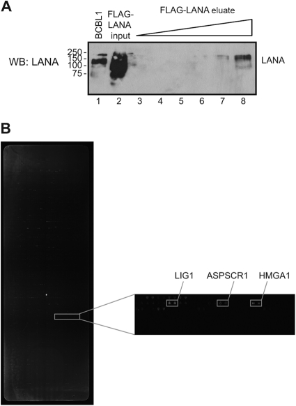
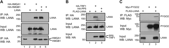
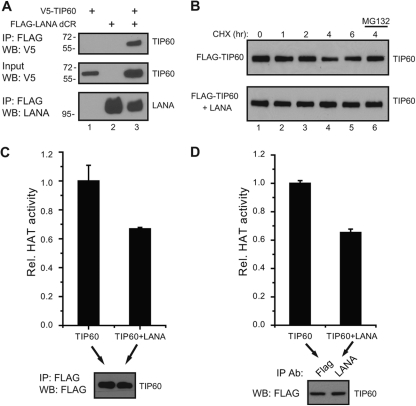
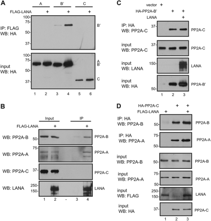
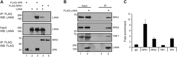
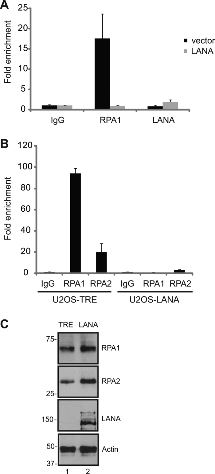
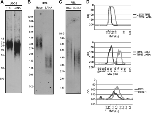
The Kaposi's sarcoma-associated herpesvirus (KSHV) LANA protein functions in latently infected cells as an essential participant in KSHV genome replication and as a driver of dysregulated cell growth. To identify novel LANA protein-cell protein interactions that could contribute to these activities, we performed a proteomic screen in which purified, adenovirus-expressed Flag-LANA protein was incubated with an array displaying 4,192 nonredundant human proteins. Sixty-one interacting cell proteins were consistently detected. LANA interactions with high-mobility group AT-hook 1 (HMGA1), HMGB1, telomeric repeat binding factor 1 (TRF1), xeroderma pigmentosum complementation group A (XPA), pygopus homolog 2 (PYGO2), protein phosphatase 2A (PP2A)B subunit, Tat-interactive protein 60 (TIP60), replication protein A1 (RPA1), and RPA2 proteins were confirmed in coimmunoprecipitation assays. LANA-associated TIP60 retained acetyltransferase activity and, unlike human papillomavirus E6 and HIV-1 TAT proteins, LANA did not reduce TIP60 stability. The LANA-bound PP2A B subunit was associated with the PP2A A subunit but not the catalytic C subunit, suggesting a disruption of PP2A phosphatase activity. This is reminiscent of the role of simian virus 40 (SV40) small t antigen. Chromatin immunoprecipitation (ChIP) assays showed binding of RPA1 and RPA2 to the KSHV terminal repeats. Interestingly, LANA expression ablated RPA1 and RPA2 binding to the cell telomeric repeats. In U2OS cells that rely on the alternative mechanism for telomere maintenance, LANA expression had minimal effect on telomere length. However, LANA expression in telomerase immortalized endothelial cells resulted in telomere shortening. In KSHV-infected cells, telomere shortening may be one more mechanism by which LANA contributes to the development of malignancy.


Figures

Fig 1
Fig 1
Expression of adenovirus 3× Flag-LANA and detection of LANA-interacting proteins on the protein array. (A) Expression of adenovirus 3× Flag-LANA. HEK 293 cells were infected with adenovirus Ad-1-LANA, and after 42 h cells were lysed, immunoprecipitated with Flag beads, and eluted with 3× Flag peptide. Cell extract from BCBL1 cells served as the LANA control (lane 1). Input (lane 2) and increasing amounts of eluted 3× Flag-LANA, dilution 1:1,000 (lane 3), 1:250 (lane 4), 1:100 (lane 5), 1:25 (lane 6), 1:10 (lane 7), and 1:5 (lane 8) were blotted with anti-LANA antibody. (B) Detection of LANA-interacting proteins on the protein array. The protein arrays were incubated with 3× Flag-LANA, followed by mouse anti-LANA antibody and Cy3-labeled goat anti-mouse antibody. One section of the protein array is presented with the gene name for some of the positive spots.
Fig 2
Fig 2
Validation of LANA interaction with HMGA1, HMGB1, TRF1, and PYGO2. (A) LANA interacts with HMGA1 and HMGB1. HEK 293T cells were transfected with expression vectors for HA-HMGA1 or HA-HMGB1 and LANA. Cell extracts were immunoprecipitated with anti-HA-conjugated beads and subjected to SDS-PAGE and Western blot analysis. (B) LANA interacts with TRF1. HEK 293T cells were transfected with expression vectors for Flag-LANA and HA-TRF1 or HA-PP5. Cell extracts were immunoprecipitated with anti-HA-conjugated beads. (C) LANA interacts with PYGO2. Cell extract from HEK 293T cells transfected with expression vectors for Flag-LANA and Myc-PYGO2 were immunoprecipitated with anti-Flag-conjugated beads.
Fig 3
Fig 3
LANA interacts with TIP60 but does not decrease its stability or abolish TIP60 acetyltransferase activity. (A) LANA interacts with TIP60. Cell extract from HEK 293T cells transfected with expression vectors for Flag-LANA dCR and V5-TIP60 were immunoprecipitated with anti-Flag-conjugated beads. (B) LANA does not reduce TIP60 stability. Flag-TIP60 was transfected alone or with a LANA expression vector, and protein extracts were prepared from cells treated with 200 μg/ml cycloheximide for the indicated times or cycloheximide plus 10 μM MG132 for 4 h. (C and D) LANA does not ablate TIP60 acetyltransferase activity. Cells were transfected with Flag-TIP60 alone or Flag-TIP60 and LANA. Cell extracts were immunoprecipitated with anti-Flag antibody to directly precipitate Flag-TIP60 (C) or with anti-LANA antibody to obtain LANA-bound Flag-TIP60 (D), and Flag-TIP60 was assayed for acetyltransferase activity. The presence of equal amounts of TIP60 in the immunoprecipitates was determined by Western blotting (bottom).
Fig 4
Fig 4
LANA interacts with PP2A. (A) LANA interacts with the B′ subunit of PP2A. Cell extract from HEK 293T cells transfected with expression vectors for Flag-LANA and HA-PP2A A, B′, or C subunits were immunoprecipitated with anti-Flag-conjugated beads. (B) LANA interacts with endogenous A and B but not C PP2A subunits. Extracts of Flag-LANA-transfected HEK 293T cells were immunoprecipitated with anti-Flag-conjugated beads and subjected to SDS-PAGE and Western blot analysis. Specific antibodies against PP2A B (top), PP2A A (second from top), and PP2A C (second from bottom) were used to detect endogenous PP2A proteins. Expression of LANA was detected with anti-LANA antibody (bottom). (C) LANA does not globally disrupt the PP2A complex. HEK 293T cells were transfected with expression vectors for HA-PP2A B′ and LANA. Total cell extracts were immunoprecipitated with anti-HA-conjugated beads and subjected to SDS-PAGE and Western blot analysis. Coimmunoprecipitated (top) and input (second from top) endogenous PP2A C were detected with anti-PP2A C antibody. (D) LANA does not globally disrupt the nuclear PP2A complex. HEK 293T cells were transfected with expression vectors for HA-PP2A C and LANA. Nuclear extracts were immunoprecipitated with anti-HA-conjugated beads and subjected to SDS-PAGE and Western blot analysis. Coimmunoprecipitated endogenous PP2A A and B were detected with anti-PP2A A and PP2A B antibodies, respectively.
Fig 5
Fig 5
RPA1 and RPA2 interact with LANA and associate with the KSHV terminal repeats. (A) LANA interacts with XPA and RPA1. Cell extract from HEK 293T cells transfected with expression vectors for Flag-XPA, Flag-RPA1, and LANA were immunoprecipitated with anti-Flag-conjugated beads. (B) LANA interacts with endogenous RPA1, RPA2, and TRF1. Extracts of Flag-LANA-transfected HEK 293T cells were immunoprecipitated with anti-Flag-conjugated beads and subjected to SDS-PAGE and Western blot analysis. Coimmunoprecipitation of endogenous RPA1 (top) RPA2 (second from top) and TRF1 (second from bottom) were detected with the respective specific antibodies. Expression of LANA was detected with anti-LANA antibody (bottom). (C) RPA1, RPA2, and XPA associate with the terminal repeats. Chromatin immunoprecipitation (ChIP) assays were performed on chromatin from BC3 cells that were immunoprecipitated with anti-RPA1, anti-RPA2, anti-TRF1, or anti-XPA antibody or control IgG. The immunoprecipitated DNA was subjected to real-time PCR using primers for the KSHV terminal repeat region. The results are presented as fold enrichment relative to control IgG. TRF1 was not associated with the terminal repeats.
Fig 6
Fig 6
LANA disrupts the association of RPA1 and RPA2 with cellular telomeres. (A) LANA disrupts the association of RPA1 with telomeres in transfected cells. ChIP assays were performed on HEK 293 cells that were transfected with a LANA expression vector. Endogenous RPA1 was immunoprecipitated and detected by real-time PCR. Fold enrichment is relative to the IgG control. (B) LANA disrupts the association of RPA1 and RPA2 with telomeres in LANA-expressing cells. ChIP assays were performed on U2OS-TRE and U2OS-LANA cell lines. (C) Expression of RPA1, RPA2, LANA, and β-actin in U2OS-TRE and U2OS-LANA cell lines as determined by Western blotting.
Fig 7
Fig 7
LANA induces telomere shortening. Telomere length analysis of DNA from U2OS-TRE and U2OS-LANA cell lines (A), from TIME-Babe and TIME-LANA cell lines (B), and from BC3 and BCBL1 cell lines (C). Cell DNA was digested with HinfI and RsaI restriction enzymes and subjected to Southern blotting. (D) Quantitation of the size distribution of telomere DNA fragments.

Similar articles

Cited by

References

    1. An J, Sun Y, Rettig MB. 2004. Transcriptional coactivation of c-Jun by the KSHV-encoded LANA. Blood 103:222–228 - PubMed
    1. Anantha RW, Borowiec JA. 2009. Mitotic crisis: the unmasking of a novel role for RPA. Cell Cycle 8:357–361 - PMC - PubMed
    1. Arnold HK, Sears RC. 2008. A tumor suppressor role for PP2A-B56alpha through negative regulation of c-Myc and other key oncoproteins. Cancer Metastasis Rev. 27:147–158 - PMC - PubMed
    1. Atanasiu C, Deng Z, Wiedmer A, Norseen J, Lieberman PM. 2006. ORC binding to TRF2 stimulates OriP replication. EMBO Rep. 7:716–721 - PMC - PubMed
    1. Awasthi S, et al. 2005. A human T-cell lymphotropic virus type 1 enhancer of Myc transforming potential stabilizes Myc-TIP60 transcriptional interactions. Mol. Cell. Biol. 25:6178–6198 - PMC - PubMed

Publication types

MeSH terms