

doi: 10.1128/JVI.06933-11.
Epub 2012 Feb 29.
Myxoma virus M064 is a novel member of the poxvirus C7L superfamily of host range factors that controls the kinetics of myxomatosis in European rabbits
Affiliations
- PMID: 22379095
- PMCID: PMC3347334
- DOI: 10.1128/JVI.06933-11
Item in Clipboard
Myxoma virus M064 is a novel member of the poxvirus C7L superfamily of host range factors that controls the kinetics of myxomatosis in European rabbits
J Virol.
2012 May.
Abstract
The myxoma virus (MYXV) carries three tandem C7L-like host range genes (M062R, M063R, and M064R). However, despite the fact that the sequences of these three genes are similar, they possess very distinctive functions in vivo. The role of M064 in MYXV pathogenesis was investigated and compared to the roles of M062 and M063. We report that M064 is a virulence factor that contributes to MYXV pathogenesis but lacks the host range properties associated with M062 and M063.
Figures
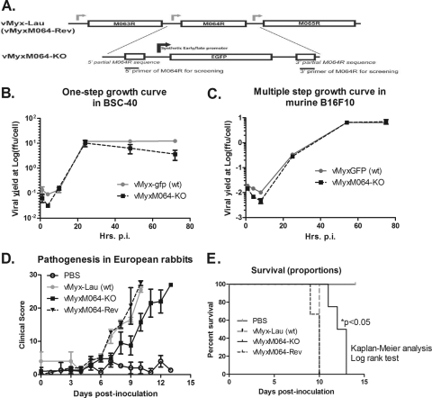
Knocking out M064R from the MYXV genome (vMyxM064R-KO) does not alter the host range tropism of the virus, but it does delay the pathogenic phenotype in vivo. (A) Construct of M064R knockout MYXV. The central region of M064R was replaced by a GFP expression cassette driven by a poxvirus synthetic early/late promoter. A revertant virus was constructed by putting back the intact sequence of M064R. (B) M064R knockout virus replicates very similarly to the wild-type virus in rabbit cells. RK-13 cells were infected at an MOI of 0.1, and cell lysates were harvested at given time points (15, 41, and 67 hours p.i.). Titration was conducted to estimate viral yields. (C) M064R knockout virus replicates similarly to the wild-type virus in murine B16F10 cells. Infection was conducted at an MOI of 0.5, and at given time points (1, 4, 8, 25, 54, and 75 h p.i.) the cell lysates were harvested for titration to estimate the viral yield. (D) M064R knockout virus showed a delayed development of myxomatosis in European rabbits. Four groups of animals were intradermally inoculated with vehicle (PBS only), 1,000 FFU of vMyx-Lau (wt), vMyxM064-KO, or vMyxM064-Rev (revertant virus). Pathogenesis was evaluated daily for each animal using a clinical score system, and the average score of each group is shown in the graph. The error bars represent the standard error of the mean (SEM). (E) M064R knockout virus infection led to a significantly delayed pathogenesis. From the same set of experiments as those shown in panel D, the survival of animals from each group was evaluated. Statistical analysis was conducted using Kaplan-Meier analysis followed by a log rank test, and a P value of <0.05 is defined as statistically significant.
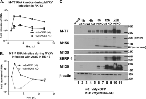
Knocking out M064R from the MYXV genome leads to a delayed expression of viral gene products during infection of rabbit cells. (A) M064R knockout virus (vMyxM064-KO) infection in rabbit cells leads to a reduced RNA transcription for viral early/late genes. RK-13 cells were infected with either wild-type MYXV (vMyxGFP) or vMyxM064-KO at an MOI of 5. At given time points (1, 2, 4, 8, and 12 h p.i.), cell lysates were harvested for RNA extraction and DNase treatment, followed by reverse transcription and then Sybr green real-time PCR to detect the transcription of an early/late gene, M-T7. The comparative CT method was used to calculate and compare the relative level of RNA. The results of the two independent experiments combined are shown. (B) The delay of vMyxM064-KO infection is at the early stage of viral gene expression. RK-13 cells were treated with AraC before and during the infection with either vMyxGFP or vMyxM064-KO. At given time points (1, 2, 4, 8, and 12 h p.i.), cell lysates were harvested and processed as described above for panel A. The results shown are representative of the results of two independent experiments. (C) The delay of viral gene expression by infection of vMyxM064-KO can be detected at the protein level. RK-13 cells were infected by either vMyxGFP (wt) or vMyxM064-KO at an MOI of 10. At given time points (1, 4, 8, 12, and 25 h p.i.), cell lysates were harvested for Western blotting. Early/late gene expression (M-T7, M156, and M135) and late gene expression (SERP-1 and M130) were compared between wt and vMyxM064-KO. The results shown are representative of the results of two independent experiments. The positions of molecular size markers in daltons are shown to the right of the gels (e.g., 50K, 50,000).
M064 is an early/late viral factor that is packaged into the progeny virions. (A) Construction of the MYXV with V5-tagged M064. A V5 tag was inserted before the stop codon of M064R, and an EGFP expression cassette driven by a vaccinia virus p11 late promoter was inserted after the V5-tagged M064R. The purity of the recombinant virus was confirmed by PCR, and this recombinant virus remains the wild-type phenotype of MYXV in vitro. (B) M064 is expressed early during viral infection, and the expressed protein stably accumulates throughout the course of infection. RK-13 cells were pretreated with AraC or not pretreated, followed by mock infection or infection with vMyxM064RV5 at an MOI of 5 in the presence or absence of AraC. At given time points (1, 2, 4, 8, 12, and 25 h p.i.), cell lysates were harvested for Western blotting. V5-tagged M064 was detected by probing with the anti-V5 antibody. Serp-1 expression was probed to show the effective AraC treatment that abolishes late gene expression. (C) M064 appears to be packaged into MYXV virions. Gradient-purified MYXV virions (0.15 optical density [OD] units for each virus) were used to coarsely separate the membrane and core components in the presence of detergent and DTT. The resulting fractions were separated on 12% SDS-polyacrylamide gels for Western blotting. V5-tagged protein was probed by anti-V5 antibody, and a known membrane component of MYXV virion, M071, was also probed as the control for a successful separation of virion core and membrane. Abbreviations: C, core component; M, membrane fraction. (D) M064 does not appear to be located in the membrane component of the purified MYXV virion. Gradient-purified MYXV virions (0.15 OD units for each virus) were used to separate only membrane and core component along with the intervened network structure which is insoluble in the absence of DTT, designated the H fraction. H+C, core fraction and H fraction that are insoluble in the absence of DTT; M, outer membrane component that is soluble in the presence of detergent while without DTT.
Similar articles
-
Disruption of M-T5, a novel myxoma virus gene member of poxvirus host range superfamily, results in dramatic attenuation of myxomatosis in infected European rabbits.J Virol. 1996 Jul;70(7):4394-410. doi: 10.1128/JVI.70.7.4394-4410.1996. J Virol. 1996. PMID: 8676463 Free PMC article.
-
M062 is a host range factor essential for myxoma virus pathogenesis and functions as an antagonist of host SAMD9 in human cells.J Virol. 2011 Apr;85(7):3270-82. doi: 10.1128/JVI.02243-10. Epub 2011 Jan 19. J Virol. 2011. PMID: 21248034 Free PMC article.
-
Comparative analysis of the complete genome sequence of the California MSW strain of myxoma virus reveals potential host adaptations.J Virol. 2013 Nov;87(22):12080-9. doi: 10.1128/JVI.01923-13. Epub 2013 Aug 28. J Virol. 2013. PMID: 23986601 Free PMC article.
-
A pox on thee! Manipulation of the host immune system by myxoma virus and implications for viral-host co-adaptation.Virus Res. 2002 Sep;88(1-2):17-33. doi: 10.1016/s0168-1702(02)00118-1. Virus Res. 2002. PMID: 12297325 Review.
-
Lessons in détente or know thy host: the immunomodulatory gene products of myxoma virus.J Biosci. 2003 Apr;28(3):273-85. doi: 10.1007/BF02970147. J Biosci. 2003. PMID: 12734406 Review.
Cited by
-
Atomic model of rabbit hemorrhagic disease virus by cryo-electron microscopy and crystallography.PLoS Pathog. 2013 Jan;9(1):e1003132. doi: 10.1371/journal.ppat.1003132. Epub 2013 Jan 17. PLoS Pathog. 2013. PMID: 23341770 Free PMC article.
-
Myxoma virus M156 is a specific inhibitor of rabbit PKR but contains a loss-of-function mutation in Australian virus isolates.Proc Natl Acad Sci U S A. 2016 Apr 5;113(14):3855-60. doi: 10.1073/pnas.1515613113. Epub 2016 Feb 22. Proc Natl Acad Sci U S A. 2016. PMID: 26903626 Free PMC article.
-
Genetic Characterization of a Recombinant Myxoma Virus in the Iberian Hare (Lepus granatensis).Viruses. 2019 Jun 7;11(6):530. doi: 10.3390/v11060530. Viruses. 2019. PMID: 31181645 Free PMC article.
-
Poxvirus Host Range Genes and Virus-Host Spectrum: A Critical Review.Viruses. 2017 Nov 7;9(11):331. doi: 10.3390/v9110331. Viruses. 2017. PMID: 29112165 Free PMC article. Review.
-
Poxviruses as Gene Therapy Vectors: Generating Poxviral Vectors Expressing Therapeutic Transgenes.Methods Mol Biol. 2019;1937:189-209. doi: 10.1007/978-1-4939-9065-8_11. Methods Mol Biol. 2019. PMID: 30706397 Free PMC article.
References
-
- Ausubel FM, et al. 1994. Current protocols in molecular biology. John Wiley and Sons, Inc., New York, NY
-
- Barrett JW, et al. 2007. Myxoma virus M063R is a host range gene essential for virus replication in rabbit cells. Virology 361:123–132 - PubMed
-
- Cameron C, et al. 1999. The complete DNA sequence of myxoma virus. Virology 264:298–318 - PubMed
Publication types
MeSH terms
Substances
Grants and funding
LinkOut - more resources
Full Text Sources
Other Literature Sources
