

Restored PB1-F2 in the 2009 pandemic H1N1 influenza virus has minimal effects in swine
- PMID: 22379102
- PMCID: PMC3347308
- DOI: 10.1128/JVI.00134-12
Restored PB1-F2 in the 2009 pandemic H1N1 influenza virus has minimal effects in swine
Abstract
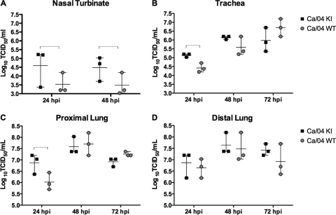
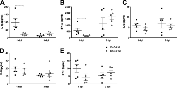
PB1-F2 is an 87- to 90-amino-acid-long protein expressed by certain influenza A viruses. Previous studies have shown that PB1-F2 contributes to virulence in the mouse model; however, its role in natural hosts-pigs, humans, or birds-remains largely unknown. Outbreaks of domestic pigs infected with the 2009 pandemic H1N1 influenza virus (pH1N1) have been detected worldwide. Unlike previous pandemic strains, pH1N1 viruses do not encode a functional PB1-F2 due to the presence of three stop codons resulting in premature truncation after codon 11. However, pH1N1s have the potential to acquire the full-length form of PB1-F2 through mutation or reassortment. In this study, we assessed whether restoring the full-length PB1-F2 open reading frame (ORF) in the pH1N1 background would have an effect on virus replication and virulence in pigs. Restoring the PB1-F2 ORF resulted in upregulation of viral polymerase activity at early time points in vitro and enhanced virus yields in porcine respiratory explants and in the lungs of infected pigs. There was an increase in the severity of pneumonia in pigs infected with isogenic virus expressing PB1-F2 compared to the wild-type (WT) pH1N1. The extent of microscopic pneumonia correlated with increased pulmonary levels of alpha interferon and interleukin-1β in pigs infected with pH1N1 encoding a functional PB1-F2 but only early in the infection. Together, our results indicate that PB1-F2 in the context of pH1N1 moderately modulates viral replication, lung histopathology, and local cytokine response in pigs.
Figures

Similar articles
-
Effects of PB1-F2 on the pathogenicity of H1N1 swine influenza virus in mice and pigs.J Gen Virol. 2017 Jan;98(1):31-42. doi: 10.1099/jgv.0.000695. J Gen Virol. 2017. PMID: 28008819 Free PMC article.
-
PB1-F2 Protein Does Not Impact the Virulence of Triple-Reassortant H3N2 Swine Influenza Virus in Pigs but Alters Pathogenicity and Transmission in Turkeys.J Virol. 2015 Oct 14;90(1):222-31. doi: 10.1128/JVI.01551-15. Print 2016 Jan 1. J Virol. 2015. PMID: 26468540 Free PMC article.
-
Strain-dependent effects of PB1-F2 of triple-reassortant H3N2 influenza viruses in swine.J Gen Virol. 2012 Oct;93(Pt 10):2204-2214. doi: 10.1099/vir.0.045005-0. Epub 2012 Jul 18. J Gen Virol. 2012. PMID: 22815274 Free PMC article.
-
Evolution and Virulence of Influenza A Virus Protein PB1-F2.Int J Mol Sci. 2017 Dec 29;19(1):96. doi: 10.3390/ijms19010096. Int J Mol Sci. 2017. PMID: 29286299 Free PMC article. Review.
-
[Swine influenza virus: evolution mechanism and epidemic characterization--a review].Wei Sheng Wu Xue Bao. 2009 Sep;49(9):1138-45. Wei Sheng Wu Xue Bao. 2009. PMID: 20030049 Review. Chinese.
Cited by
-
Influenza A virus PB1-F2 protein expression is regulated in a strain-specific manner by sequences located downstream of the PB1-F2 initiation codon.J Virol. 2013 Oct;87(19):10687-99. doi: 10.1128/JVI.01520-13. Epub 2013 Jul 24. J Virol. 2013. PMID: 23885074 Free PMC article.
-
The ubiquitination of the influenza A virus PB1-F2 protein is crucial for its biological function.PLoS One. 2015 Apr 13;10(4):e0118477. doi: 10.1371/journal.pone.0118477. eCollection 2015. PLoS One. 2015. PMID: 25866881 Free PMC article.
-
Non-avian animal reservoirs present a source of influenza A PB1-F2 proteins with novel virulence-enhancing markers.PLoS One. 2014 Nov 4;9(11):e111603. doi: 10.1371/journal.pone.0111603. eCollection 2014. PLoS One. 2014. PMID: 25368997 Free PMC article.
-
Influenza A virus PB1-F2 protein prolongs viral shedding in chickens lengthening the transmission window.J Gen Virol. 2016 Oct;97(10):2516-2527. doi: 10.1099/jgv.0.000584. Epub 2016 Aug 23. J Gen Virol. 2016. PMID: 27558742 Free PMC article.
-
Effects of the PA-X and PB1-F2 Proteins on the Virulence of the 2009 Pandemic H1N1 Influenza A Virus in Mice.Front Cell Infect Microbiol. 2019 Sep 3;9:315. doi: 10.3389/fcimb.2019.00315. eCollection 2019. Front Cell Infect Microbiol. 2019. PMID: 31552197 Free PMC article.
References
-
- Brockmeier SL, et al. 2009. Adenovirus-mediated expression of interferon-alpha delays viral replication and reduces disease signs in swine challenged with porcine reproductive and respiratory syndrome virus. Viral Immunol. 22:173–180 - PubMed
Publication types
MeSH terms
Substances
Grants and funding
LinkOut - more resources
Full Text Sources
Miscellaneous
