. 2012 Jun;81(6):759-69.
doi: 10.1124/mol.112.077792. Epub 2012 Feb 29.

Pharmacological chaperoning of nicotinic acetylcholine receptors reduces the endoplasmic reticulum stress response

Affiliations

Pharmacological chaperoning of nicotinic acetylcholine receptors reduces the endoplasmic reticulum stress response

Rahul Srinivasan et al. Mol Pharmacol. 2012 Jun.

Abstract

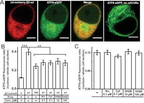
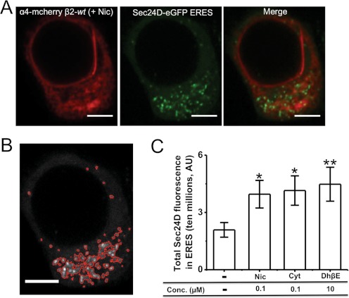
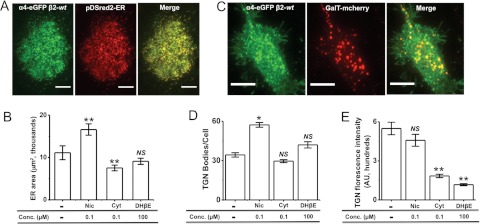
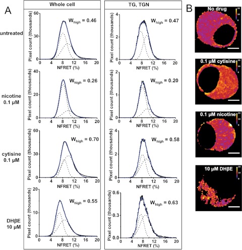
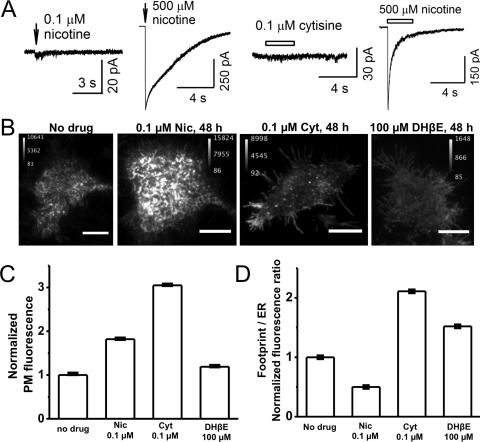
We report the first observation that endoplasmic reticulum (ER) stress and the unfolded protein response (UPR) can decrease when a central nervous system drug acts as an intracellular pharmacological chaperone for its classic receptor. Transient expression of α4β2 nicotinic receptors (nAChRs) in Neuro-2a cells induced the nuclear translocation of activating transcription factor 6 (ATF6), which is part of the UPR. Cells were exposed for 48 h to the full agonist nicotine, the partial agonist cytisine, or the competitive antagonist dihydro-β-erythroidine; we also tested mutant nAChRs that readily exit the ER. Each of these four manipulations increased Sec24D-enhanced green fluorescent protein fluorescence of condensed ER exit sites and attenuated translocation of ATF6-enhanced green fluorescent protein to the nucleus. However, we found no correlation among the manipulations regarding other tested parameters [i.e., changes in nAChR stoichiometry (α4(2)β2(3) versus α4(3)β2(2)), changes in ER and trans-Golgi structures, or the degree of nAChR up-regulation at the plasma membrane]. The four manipulations activated 0 to 0.4% of nAChRs, which shows that activation of the nAChR channel did not underlie the reduced ER stress. Nicotine also attenuated endogenously expressed ATF6 translocation and phosphorylation of eukaryotic initiation factor 2α in mouse cortical neurons transfected with α4β2 nAChRs. We conclude that, when nicotine accelerates ER export of α4β2 nAChRs, this suppresses ER stress and the UPR. Suppression of a sustained UPR may explain the apparent neuroprotective effect that causes the inverse correlation between a person's history of tobacco use and susceptibility to developing Parkinson's disease. This suggests a novel mechanism for neuroprotection by nicotine.


Figures

Fig. 1.
Fig. 1.
Each of the four manipulations decreases ATF6 translocation in Neuro-2a cells. A, representative confocal images of Neuro-2a cells expressing α4-mcherryβ2- wt nAChRs plus ATF6-eGFP or only ATF6-eGFP. Scale bars, 10 μm. B, ATF6-eGFP fluorescence intensity ratios (nucleus/whole cell section). Treatment conditions and transfected subunits are indicated on the x-axis. DM, double-mutant β2enhanced-ER-export subunit. C, ATF6-eGFP fluorescence intensity ratios (nucleus/whole cell section) in the absence of coexpressed nAChRs. Drug treatments are indicated on the x-axis. The p values are based on a 2-tailed t test. **, p < 0.01; ***, p < 0.001. Data were obtained from 30 to 40 cells imaged for each condition. Nic, nicotine; Cyt, cytisine.
Fig. 2.
Fig. 2.
Nicotine inhibits ATF6 translocation to the nucleus and eIF2α phosphorylation in mouse cortical neurons. A, representative confocal images of mouse cortical neurons expressing α4-mcherryβ2-wt, with immunostaining for endogenous ATF6, peIF2α, or total eIF2α (scale bars, 10 μm). B, fluorescence intensity ratios for endogenously expressed ATF6, with and without 0.1 μM nicotine (Nic) treatment for 24 h. C, nuclear fluorescence intensity for peIF2α, with and without 0.1 μM nicotine treatment for 24 h. D, fluorescence intensity for total eIF2α. Bars show nicotine treatment conditions. All neurons in B, C, and D expressed transfected α4-mcherry plus β2-wt subunits. The subcellular compartments imaged are indicated on the x-axis. The p values are based on a 2-tailed t test. *, p < 0.05; **, p < 0.01. Data were obtained from 20 to 30 cells imaged for each condition.
Fig. 3.
Fig. 3.
Exposure to all three nicotinic ligands increases the formation of ERES. A, representative confocal images of a Neuro-2a cell expressing α4-mcherryβ2-wt nAChRs and Sec24D-eGFP (ERES marker). Scale bars, 10 μm. B, image of the same cell with ERES demarcated for quantification. C, quantification of total Sec24D fluorescence in ERES per cell. Drug treatment conditions are indicated on the x-axis. Error bars indicate S.E.M. The p values are based on a 2-tailed t test. *, p < 0.05; **, p < 0.01. The graphs present total Sec24D fluorescence of ERES per cell, averaged for 30 to 40 cells imaged for each condition. Nic, nicotine; Cyt, cytisine.
Fig. 4.
Fig. 4.
The three nicotinic ligands have diverse effects on ER and TGN architecture. A, representative TIRFM images of a cell coexpressing α4-eGFPβ2-wt nAChRs and pDSred2-ER marker. The merged image shows nearly complete colocalization of fluorescence from nAChRs and DSred2-ER. Scale bars, 10 μm. B, quantification of average ER area determined by using DSred2-ER fluorescence. Drug treatments are indicated on the x-axis. C, representative TIRFM images of a cell expressing α4-eGFPβ2-wt nAChRs and GalT-mcherry. The merged image shows colocalization of GalT-mcherry with nAChRs. Scale bars, 10 μm. D, quantification of the number of TGN bodies observed by using GalT-mcherry under each treatment condition, as indicated on the x-axis. E, quantification of the average TGN body fluorescence intensity observed by using GalT-mcherry under each treatment condition, as indicated on the x-axis. Error bars indicate S.E.M. The p values are based on a 2-tailed t test. *, p < 0.05; **, p < 0.01. Data were obtained from 30 to 40 cells imaged for each condition. Nic, nicotine; Cyt, cytisine; NS, not significant.
Fig. 5.
Fig. 5.
The three nicotinic ligands have diverse effects on α4β2 nAChR stoichiometry. A, NFRET measurements were made by using α4-mcherry and β2-eGFP subunits transfected into Neuro-2a cells. Columns indicate the ROIs (whole cell or TG, TGN), and rows indicate drug treatment conditions. For each graph, the blue curve is the overall fit and dashed Gaussian curves are individual fits. Whigh, corresponding to the α43β22 stoichiometry, is shown for each graph. B, representative NFRET images of Neuro-2a cells. Drug treatments are indicated for each cell. Calibration bars are from zero to 20%. Scale bars, 5 μm.
Fig. 6.
Fig. 6.
The three nicotinic ligands have diverse effects on PM-localized α4β2 nAChRs. A, representative traces showing whole-cell currents induced by puffs of 0.1 and 500 μM nicotine or 0.1 μM cytisine and 500 μM nicotine. B, representative TIRFM images of cells expressing α4-eGFPβ2-wt nAChRs with treatment with the indicated drugs for 48 h. Scale bars, 10 μm. C, bar graph showing normalized PM fluorescence intensity from TIRFM images, normalized as described in Materials and Methods. Drug treatment conditions are shown on the x-axis. Error bars indicate 99% confidence intervals, which show that all differences are highly significant. D, footprint/ER ratios from TIRFM images. Drug treatment conditions are shown on the x-axis. Error bars indicate relative S.E. Data were obtained from 30 to 40 cells imaged for each condition. Nic, nicotine; Cyt, cytisine.

References

    1. Ahmadi FA, Grammatopoulos TN, Poczobutt AM, Jones SM, Snell LD, Das M, Zawada WM. (2008) Dopamine selectively sensitizes dopaminergic neurons to rotenone-induced apoptosis. Neurochem Res 33:886–901 - PubMed
    1. Azam L, Winzer-Serhan UH, Chen Y, Leslie FM. (2002) Expression of neuronal nicotinic acetylcholine receptor subunit mRNAs within midbrain dopamine neurons. J Comp Neurol 444:260–274 - PubMed
    1. Bommiasamy H, Back SH, Fagone P, Lee K, Meshinchi S, Vink E, Sriburi R, Frank M, Jackowski S, Kaufman RJ, et al. (2009) ATF6α induces XBP1-independent expansion of the endoplasmic reticulum. J Cell Sci 122:1626–1636 - PMC - PubMed
    1. Buisson B, Bertrand D. (2001) Chronic exposure to nicotine upregulates the human α4β2 nicotinic acetylcholine receptor function. J Neurosci 21:1819–1829 - PMC - PubMed
    1. Buisson B, Bertrand D. (2002) Nicotine addiction: the possible role of functional upregulation. Trends Pharmacol Sci 23:130–136 - PubMed

Publication types

MeSH terms

LinkOut - more resources