. 2012 Mar 15;26(6):567-80.
doi: 10.1101/gad.183251.111. Epub 2012 Feb 29.

Simulated body temperature rhythms reveal the phase-shifting behavior and plasticity of mammalian circadian oscillators

Affiliations

Simulated body temperature rhythms reveal the phase-shifting behavior and plasticity of mammalian circadian oscillators

Camille Saini et al. Genes Dev. .

Abstract

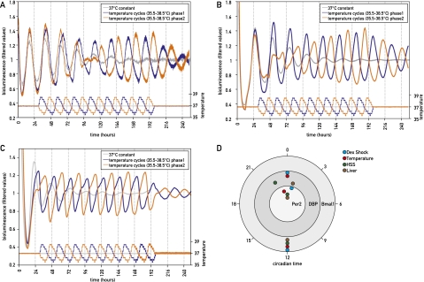
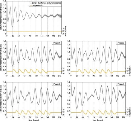
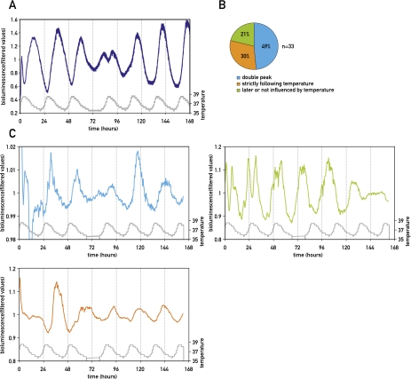
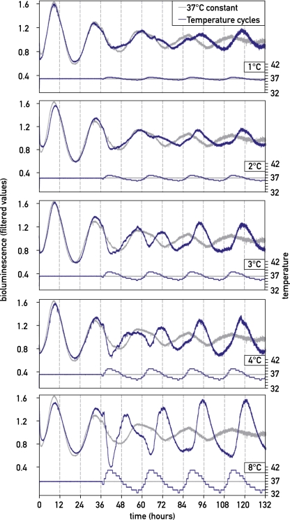
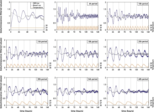
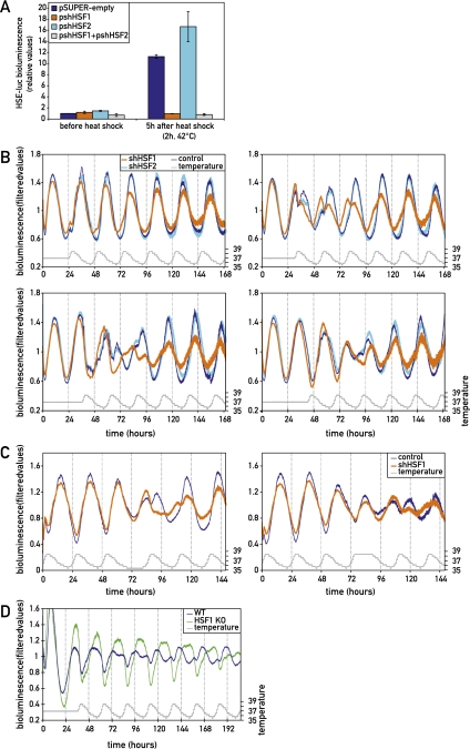
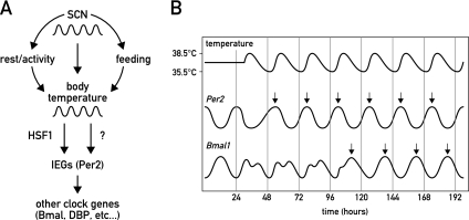
The circadian pacemaker in the suprachiasmatic nuclei (SCN) of the hypothalamus maintains phase coherence in peripheral cells through metabolic, neuronal, and humoral signaling pathways. Here, we investigated the role of daily body temperature fluctuations as possible systemic cues in the resetting of peripheral oscillators. Using precise temperature devices in conjunction with real-time monitoring of the bioluminescence produced by circadian luciferase reporter genes, we showed that simulated body temperature cycles of mice and even humans, with daily temperature differences of only 3°C and 1°C, respectively, could gradually synchronize circadian gene expression in cultured fibroblasts. The time required for establishing the new steady-state phase depended on the reporter gene, but after a few days, the expression of each gene oscillated with a precise phase relative to that of the temperature cycles. Smooth temperature oscillations with a very small amplitude could synchronize fibroblast clocks over a wide temperature range, and such temperature rhythms were also capable of entraining gene expression cycles to periods significantly longer or shorter than 24 h. As revealed by genetic loss-of-function experiments, heat-shock factor 1 (HSF1), but not HSF2, was required for the efficient synchronization of fibroblast oscillators to simulated body temperature cycles.


Figures

Figure 1.
Figure 1.
Synchronization of cultured fibroblast clocks by simulated body temperature fluctuations. NIH3T3/Bmal1-luc cells (A), NIH3T3/Dbp-luc cells (B), or immortalized tail fibroblasts from PER2::luciferase knock-in mice (C) were exposed to temperature cycles resembling body temperature rhythms measured telemetrically in mice (35.5°C–38.5°C) with opposite phases (blue and orange) and then released into a constant temperature of 37°C. Parallel cell populations were continuously kept at 37°C temperature (gray). All bioluminescence data were filtered by a moving average transformation (see the Materials and Methods). (D) After the synchronization of mouse fibroblasts by simulated body temperature cycles (red), the phase relationship was conserved for the expression of Bmal1, Dbp, and Per2. The differences in phase angles were similar to those observed in cells transiently synchronized by a short treatment with 50% horse serum (green) or dexamethasone (light blue) or those determined in the liver of intact animals (brown).
Figure 2.
Figure 2.
The resynchronization kinetics of circadian Bmal1-luciferase expression by simulated body temperature cycles depend on the old phase. NIH3T3/Bmal1-luc cells were transiently synchronized with horse serum and then subjected to temperature cycles starting 24, 30, 36, or 42 h after the serum treatment (phases 1, 2, 3, and 4). The top left panel shows bioluminescence from a cell population kept at constant temperature. The next four panels show the responses of cells subjected to temperature phases 1, 2, 3, and 4. All bioluminescence data were filtered by moving average transformation.
Figure 3.
Figure 3.
Bioluminescence microscopy of individual cells reveals different sensitivities to temperature cycles. (A) Bioluminescence monitoring of Bmal1-luciferase expression in NIH3T3 fibroblasts subjected to three temperature cycles, followed by a 12-h phase shift (gray curve) at the level of the whole population (dark blue) using photomultiplier tube technology for data acquisition, or at the single-cell level (three panels in C) using a LV200 luminoview microscope (Olympus) equipped with an EM-CCD cooled camera (Hamamatsu Photonics) (see the Materials and Methods). Three different patterns were observed in individual cells. The occurrence of each of them is quantified in B, and a representative example of each of them is shown in C. In the first case (turquoise), the cell was entrained by the initial phase, and this phase entered in conflict with the new phase, giving rise to a “double-peak phenotype.” In the second case (green), the cell kept its initial phase without responding to the new phase of the temperature cycle. In the third case (orange), the cell strictly followed the phase of the temperature cycles and immediately switched to the new phase. All bioluminescence data were filtered by moving average transformation (see the Materials and Methods).
Figure 4.
Figure 4.
Circadian clocks can be partially synchronized by temperature fluctuations with very small amplitudes. Circadian gene expression was transiently synchronized by a horse serum shock, and cells were exposed to temperature oscillations with amplitudes differing by 1°C, 2°C, 3°C, 4°C, or 8°C and with a phase opposite to that imposed by the serum shock (corresponding to phase 3 in Fig. 2). Note that all temperature cycles were able to synchronize at least a fraction of NIH3T3/Bmal1-luc cells. All bioluminescence data were filtered by moving average transformation.
Figure 5.
Figure 5.
Circadian gene expression adapts to widely different T-cycles of simulated body temperature rhythms. NIH3T3 fibroblasts transfected with a Bmal1-luciferase reporter (blue) or a CMV-luciferase reporter (gray; used as a noncircadian control) were kept in constant temperature (first panel) or were exposed to temperature T-cycles of 35.5°C–38.5°C with period lengths of 6, 10, 14, 18, 24, 30, 34, or 40 h (as indicated). All bioluminescence data were filtered by moving average transformation.
Figure 6.
Figure 6.
HSF1 participates in the synchronization of circadian clocks by simulated body temperature cycles. (A) The response of an HSE-luciferase reporter gene to a heat shock (2 h, 42°C) is completely abolished when NIH3T3 cells are cotransfected with a plasmid (pshHSF1) encoding a shRNA targeting Hsf1 mRNA (orange). In contrast, cotransfections with a plasmid (pshHSF2) specifying a shRNA targeting Hsf2 mRNA (light blue) or an empty control plasmid (pSUPER) (blue) had no effect on the heat-induced expression of HSE-luciferase. Bioluminescence intensities were determined before and 5 h after the heat shock and were plotted against the background bioluminescence levels determined for pSUPER before the heat shock. The response of circadian Bmal1-luciferase reporter to simulated body temperature cycles with a phase opposite to the old phase (B) or to a phase inversion in temperature cycles (C) was delayed when NIH3T3 cells were cotransfected with pshHSF1 (orange). In contrast, the cotransfection of cells with pshHSF2 (light blue) or pSUPER (blue) had little effect on the phase-shifting kinetics of circadian Bmal1-luciferase expression. (D) Primary tail fibroblasts from Hsf1 knockout mice that stably express a Bmal1-luciferase reporter (green) were more slowly synchronized by simulated body temperature fluctuations of a contradictory phase when compared with tail fibroblasts from wild-type mice (blue). In B and D, cells were synchronized with horse serum prior to the initiation of bioluminescence monitoring. Note also that the kinetics of adaptation to the new phase depended on the cell type (NIH3T3 vs. primary tail fibroblasts) and on whether the reporter gene was transiently transfected or stably integrated. All bioluminescence data were filtered by moving average transformation.
Figure 7.
Figure 7.
Model for the integration of body temperature rhythms in the synchronization of peripheral oscillators. (A) In mammals, the SCN regulates virtually all clock outputs, including rest/activity cycles, feeding/fasting cycles, and temperature rhythms. In peripheral cell types, elevated temperature induces HSF1 activity, which, in cooperation with other temperature-sensitive regulators, promotes the expression of immediate early genes (IEGs) such as Per2. In turn, the IEGs phase-reset other clock genes such as Bmal1 or Dbp. A direct action of temperature on the activity and stability of clock components may also occur. (B) Representative expression kinetics of Per2 versus Bmal1 in response to an altered body temperature rhythm. While Per2 expression immediately adapts to the temperature stimulus, Bmal1 expression requires few days to oscillate in resonance with the newly imposed phase.

Similar articles

Cited by

References

    1. Abraham U, Granada AE, Westermark PO, Heine M, Kramer A, Herzel H 2011. Coupling governs entrainment range of circadian clocks. Mol Syst Biol 6: 438 doi: 10.1038/msb.2010.92 - PMC - PubMed
    1. Akashi M, Nishida E 2000. Involvement of the MAP kinase cascade in resetting of the mammalian circadian clock. Genes Dev 14: 645–649 - PMC - PubMed
    1. Akhtar RA, Reddy AB, Maywood ES, Clayton JD, King VM, Smith AG, Gant TW, Hastings MH, Kyriacou CP 2002. Circadian cycling of the mouse liver transcriptome, as revealed by cDNA microarray, is driven by the suprachiasmatic nucleus. Curr Biol 12: 540–550 - PubMed
    1. Albrecht U, Sun ZS, Eichele G, Lee CC 1997. A differential response of two putative mammalian circadian regulators, mper1 and mper2, to light. Cell 91: 1055–1064 - PubMed
    1. Asher G, Reinke H, Altmeyer M, Gutierrez-Arcelus M, Hottiger MO, Schibler U 2010. Poly(ADP-ribose) polymerase 1 participates in the phase entrainment of circadian clocks to feeding. Cell 142: 943–953 - PubMed

Publication types

LinkOut - more resources