. 2011 Mar;28(1):48-62.
doi: 10.1055/s-0031-1273940.

Bronchial artery embolization for hemoptysis

Affiliations

Bronchial artery embolization for hemoptysis

David R Sopko et al. Semin Intervent Radiol. 2011 Mar.

Abstract

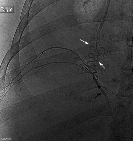
Bronchial artery angiography with embolization has become a mainstay in the treatment of hemoptysis. Major complications are rare and immediate clinical success defined as cessation of hemorrhage ranges in most series from 85% to 100%, although recurrence of hemorrhage ranges from 10% to 33%. Bronchial artery embolization offers a minimally invasive procedure for even the most compromised patient serving as first-line treatment for hemorrhage as well as providing a bridge to more definitive medical or surgical intervention focused upon the etiology of the hemorrhage. The aim of this article is to summarize the etiologies, pathophysiology, and the diagnostic and management strategies of hemoptysis as related to bronchial artery embolization. In addition, the techniques of arteriography and embolization as well as associated procedural outcomes and complications are delineated.

Keywords: Hemoptysis; angiography; arterial embolization; bronchial artery.


Figures

Figure 1
Figure 1
The four most prevalent patterns of bronchial artery anatomy. Type I: single right bronchial artery via intercostobronchial trunk (ICBT), paired left bronchial arteries. Type II: single right bronchial artery via ICBT, single left bronchial artery. Type III: paired right bronchial arteries with one from ICBT, paired left bronchial arteries. Type IV: paired right bronchial arteries with one from ICBT, solitary left bronchial artery.
Figure 2
Figure 2
(A) A 24-year-old man undergoing spinal angiography for hemorrhage. Thoracic aortogram shows single bronchial artery (arrow) supplying left and right sides. (B) Selective angiogram of the single bronchial artery from the aortogram shows normal bronchial artery distribution. A single trunk (arrowhead) gives rise to a right and left bronchial artery (arrows). Note the relatively small caliber of the normal arteries. (C) Normal bronchial angiogram in a 67-year-old man with right lung mass and hemoptysis following bronchoscopy. Angiography demonstrates a common origin (arrowhead) of the left (arrow) and right (arrow) bronchial arteries. Note the small caliber of the normal vessels. Given the patient's history, embolization with 150–250-μm polyvinyl alcohol particles was performed despite normal vessel appearance.
Figure 3
Figure 3
A 66-year-old woman with sarcoidosis and hemoptysis. Only a very small right bronchial artery found on selective catheterization and aortography. Celiac artery arteriography showed mildly enlarged right phrenic artery. Subselective angiogram of the right phrenic artery (black arrow) shows arterial flow (white arrows) to the poorly aerated right lung base. This was successfully embolized using 250–350-μm polyvinyl alcohol particles.
Figure 4
Figure 4
A 54-year-old man with known bronchiectasis and prior right upper lobe lung resection now presenting with aspergillosis and hemoptysis. (A) Early-phase arteriogram of the right bronchial artery (black arrow) showing abnormal hypervascularity (white arrows) of the diseased partially collapsed right lower lobe. (B) Late-phase arteriogram of the right bronchial artery showing venous drainage via the pulmonary veins (arrows). This bronchial artery was successfully embolized with 355–500-μm polyvinyl alcohol particles.
Figure 5
Figure 5
(A) A 24-year-old man undergoing spinal angiography for hemorrhage, same patient as Fig. 2A. Injection of the left T12 intercostal artery demonstrates a prominent normal anterior spinal artery (artery of Adamkiewicz) (arrows). (B) A 24-year-old woman with cystic fibrosis and hemoptysis. Injection of the right supreme intercostal artery (black arrowhead) demonstrates a large, abnormal bronchial artery (white arrow) designating this as an intercostobronchial trunk. Note supply to the anterior spinal artery from the supreme intercostal arterial supply (black arrows). Embolization was performed in this patient beyond the origin of the supreme intercostal artery with the microcatheter placed at the level of the white arrow (see Fig. 8). Care was taken not to reflux particles into the supreme intercostal artery distribution (white arrowheads).
Figure 6
Figure 6
A 52-year-old female with sarcoidosis, bronchiectasis, and cavitation with mycetoma. She presents with recurrent hemoptysis. (A) Aortogram (frontal view) shows enlarged left T6 intercostal artery (arrow) providing a tortuous hypervascular supply to abnormal tissue. In addition, a previously embolized left bronchial artery (black arrowhead) that has partially recanalized is also seen. Supply is also noted from a left supreme intercostal artery (white arrowhead). The patient has undergone previous successful coil embolization on the right (white arrow). Aortography nicely provides a working roadmap as it is useful to visualize bronchial arteries and collateral supply including supply from the intercostal arteries as shown here. (B) Left sixth intercostal arteriogram in the early phase shows collateral supply (black arrows) to abnormal left lung tissue. Note prior embolization coils (white arrows). (C) Left sixth intercostal arteriogram in a later phase shows branches of the pulmonary artery (arrows) consistent with shunting. As the shunting is through abnormal tissue with small vessels, this intercostal artery was successfully and safely embolized using larger (355–500 μm) polyvinyl alcohol particles.
Figure 7
Figure 7
(A) 45-year-old man with sarcoidosis and aspergilloma presenting with hemoptysis. Single image from axial computed tomography shows pleural thickening (black arrow) and aspergilloma (white arrowhead), which was the etiology of this patient's hemoptysis. (B) Right third intercostal artery (arrow) gives rise to collaterals (arrowheads) supplying the aspergilloma. (C) Right third intercostal following successful embolization with 150–250-μm polyvinyl alcohol particles via a microcatheter (arrow). Note the lack of blood flow to the area of the mycetoma (arrowhead).
Figure 8
Figure 8
(A) A 24-year-old woman with cystic fibrosis and hemoptysis, same patient as Fig. 5B. Chest radiograph shows bilateral opacities in this patient with cystic fibrosis. (B) Injection of the left supreme intercostal artery shows the enlarged bronchial artery (arrow). (C) A microcatheter (arrowhead) was placed beyond the intercostal branch, which contributes arterial supply to the anterior spinal artery (see Fig. 5B), and embolization was successfully performed using large (1000–1180 μm) polyvinyl alcohol particles. Larger particles were used to prevent migration into spinal artery supply should accidental reflux transpire, although care was taken not to reflux into the intercostal artery. (D) Postembolization angiogram of the right supreme intercostobronchial trunk. Note the very slow flow in the bronchial artery (arrow) and its distal branches (black arrowheads). Microcatheter tip is in the intercostobronchial trunk (white arrowhead). Note the excellent filling of the distal supreme intercostal artery, which supplied the anterior spinal artery in the lower cervical/upper thoracic region (Fig. 5B). Patient was neurologically intact following the procedure.
Figure 9
Figure 9
(A) A 12-year-old woman with Lennox-Gastaut syndrome and history of recurrent hemoptysis with multiple previous embolization procedures. As this patient had undergone multiple prior bronchial embolization procedures, pulmonary angiogram was performed to exclude this arterial circulation as a source. It is normal with no evidence for a bleeding site. (B) Angiogram via a microcatheter (white arrowhead) of an enlarged collateral branch of the left thyrocervical artery shows collateral filling (black arrows) around and through the coils placed from a previous embolization. Proximal embolization such as with coils can often lead to this situation. (C) Embolization successfully performed via the microcatheter (white arrowhead) using 355–500 μm polyvinyl alcohol particles resulting in slow flow in the main trunk (black arrow) and no flow distally (black arrowheads).

Similar articles

Cited by

References

    1. Haponik E F, Fein A, Chin R. Managing life-threatening hemoptysis: has anything really changed? Chest. 2000;118(5):1431–1435. - PubMed
    1. Shigemura N, Wan I Y, Yu S C, et al. Multidisciplinary management of life-threatening massive hemoptysis: a 10-year experience. Ann Thorac Surg. 2009;87(3):849–853. - PubMed
    1. Wyngaarden J B, Smith L H, Bennett J C. Cecil Textbook of Medicine. 19th ed. Philadelphia: WB Saunders; 1992. p. 370.
    1. Crocco J A, Rooney J J, Fankushen D S, DiBenedetto R J, Lyons H A. Massive hemoptysis. Arch Intern Med. 1968;121(6):495–498. - PubMed
    1. Ferris E J. Pulmonary hemorrhage. Vascular evaluation and interventional therapy. Chest. 1981;80(6):710–714. - PubMed