. 2011 Mar;28(1):63-74.
doi: 10.1055/s-0031-1273941.

Thoracic duct embolization for chylous leaks

Affiliations

Thoracic duct embolization for chylous leaks

Eric Chen et al. Semin Intervent Radiol. 2011 Mar.

Abstract

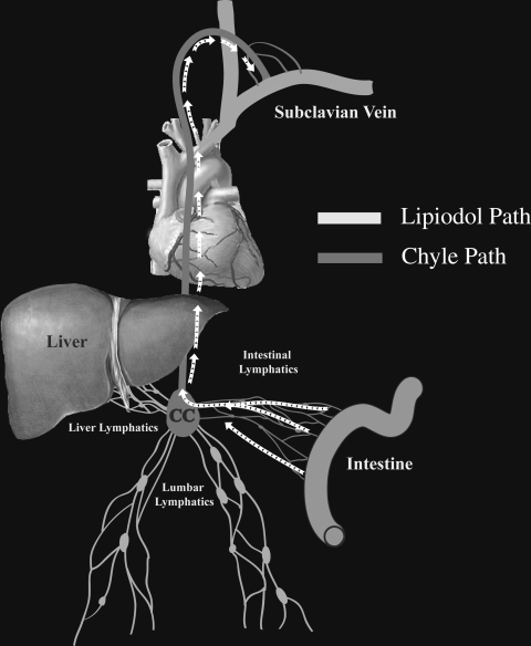
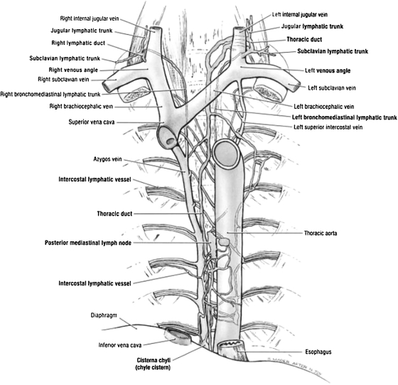
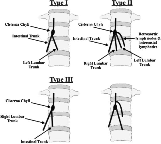
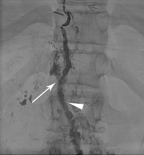
Chylous leaks, such as chylothorax and chylopericardium, are uncommon effusions resulting from the leakage of intestinal lymphatic fluid from the thoracic duct (TD) and its tributaries, or intestinal lymphatic ducts. The cause can be either traumatic (thoracic surgery) or nontraumatic (idiopathic, malignancy). Treatment has traditionally consisted of dietary modification (nonfat diet) and/or surgery (TD ligation, pleurodesis). Thoracic duct embolization (TDE) has become a viable treatment alternative due to it high success rate and minimal complications. In this article, the authors describe the etiologies of chylothorax, patient population, outcomes, and long-term follow-up of TDE patients. Relevant lymphatic anatomy and physiology are reviewed, with special attention paid to the formation of the duct by tributaries at the cisterna chyli (CC). The technique of TDE is outlined, including bilateral pedal lymphangiography, TD cannulation, and embolic agents used for the procedure.

Keywords: Thoracic duct embolization; chyle leak; chylothorax.


Figures

Figure 1
Figure 1
Schematic representation of the chyle pathway. CC, Cisterna chyli.
Figure 2
Figure 2
Anatomic rendering of the thoracic duct. (Adapted from Agur AMR, Dalley AF, Grant JCB. Grant's Atlas of Anatomy. 11th ed. Philadelphia: Lippincott Williams & Wilkins; 2005.)
Figure 3
Figure 3
Schematic representation of the major anatomic variants of the thoracic duct based on embryologic development and the rate of their occurrence observed on magnetic resonance ductography. (Reprinted with permission from Okuda et al. Magnetic resonance-thoracic ductography: imaging aid for thoracic surgery and thoracic duct depiction based on embryological considerations. Gen Thorac Cardiovasc Surg 2009;57(12):640–646.)
Figure 4
Figure 4
Schematic representation of the five major anatomic variants of the thoracic duct and the rate of their occurrence observed in postmortem study. (Reprinted with permission from Kausel et al. Anatomic and pathologic studies of the thoracic duct. J Thorac Surg 1957;34(5):631–641.)
Figure 5
Figure 5
Computed tomography image of the retrocrural location of the cisterna chyli following lymphangiogram (arrow).
Figure 6
Figure 6
Schematic representation of the anatomic variation of the tributaries of the cisterna chyli. (Reprinted with permission from Loukas et al. Cisterna chyli: a detailed anatomic investigation. Clin Anat 2007;20(6):683–688.)
Figure 7
Figure 7
Technical steps of thoracic duct (TD) embolization. (A) Fluoroscopic image of the access of the cisterna chyli with a 21-gauge needle (arrowhead). (B) Fluoroscopic imaging of the V-18 wire advanced into the TD (arrow). (C) Injection of the contrast into the TD through the catheter (arrow) to identify the chylous leak (arrowhead). (D) Final fluoroscopy image after embolization of the TD with microcoils (arrow) and Truefill glue.
Figure 8
Figure 8
Fluoroscopic images of four lymphangiography patterns of the thoracic duct (TD) nontraumatic chylous effusion population. (A) Distal part of normal TD (arrow) and the termination of the TD at the subclavian vein (arrowhead). (B) Injection of the contrast agent into TD via microcatheter (arrow) demonstrated complete occlusion of the TD with development of multiple collaterals (arrowhead). (C) Complete occlusion of the caudal lymphatic flow at the level of the midabdomen (black arrow). Patient with the history of previously treated lymphoma. (D) Leakage of the contrast from the cisterna chyli (white arrow) in patient post unsuccessful TD ligation. Note distended cisterna chyli (arrowhead).
Figure 9
Figure 9
Lymphangiographic image demonstrating contrast extravasation (arrow) from the hole created by previous access of the cisterna chyli (CC). Note the contrast in the CC from the first thoracic duct embolization (arrowhead).

References

    1. Dunn R P. Primary chylopericardium: a review of the literature and an illustrated case. Am Heart J. 1975;89(3):369–377. - PubMed
    1. Itkin M, Swe N M, Shapiro S E, Shrager J B. Spontaneous chylopericardium: delineation of the underlying anatomic pathology by CT lymphangiography. Ann Thorac Surg. 2009;87(5):1595–1597. - PubMed
    1. Doerr C H, Allen M S, Nichols F C, III, Ryu J H. Etiology of chylothorax in 203 patients. Mayo Clin Proc. 2005;80(7):867–870. - PubMed
    1. Cerfolio R J, Allen M S, Deschamps C, Trastek V F, Pairolero P C. Postoperative chylothorax. J Thorac Cardiovasc Surg. 1996;112(5):1361–1365. discussion 1365–1366. - PubMed
    1. Dougenis D, Walker W S, Cameron E W, Walbaum P R. Management of chylothorax complicating extensive esophageal resection. Surg Gynecol Obstet. 1992;174(6):501–506. - PubMed