

Ophthalmic considerations in cleft lip and palate patients
- PMID: 22379315
- PMCID: PMC3177494
- DOI: 10.1007/s12663-010-0058-z
Ophthalmic considerations in cleft lip and palate patients
Abstract
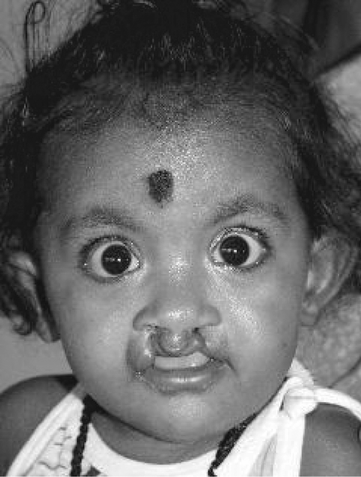
Cleft lip and palate (CL and P) represents the second most frequently occurring congenital deformity after clubfoot deformity. CL and P could be associated with many other structural abnormalities of the adjacent vital structures of the face. In this study, an attempt was made to identify the abnormalities of the ocular structures seen in patients with isolated CL and P as well as in those with syndromic CL and P. Of the 322 patients with cleft lip and palate screened, 27 (8.3%) had ocular abnormalities. Totally 47 ocular defects were identified in 27 patients. Abnormalities of the eyelid were the commonest accounting for 22% of the total defects (11/47), which includes symblepharon, ectropion, lid colobomas, euryblepharon and ptosis. Second commonest abnormality was squint 8/47 (17%) followed by orbital defects 8/47 (17%) (Telecanthus and Hypertelorism). Abnormalities of the nasolacrimal apparatus 3/47 (6%), refractive errors 7/47 (15%), dermoids 4/47 (6%), cataract 2/47 (4%) and retinal colobomas 2/47 (4%) constituted the rest. Thus, children with cleft lip and palate should be assessed as soon as possible after birth by a multidisciplinary team involving the Pediatrician, Ophthalmologist and specialists from Maxillofacial, ENT and Plastic surgery. The medical problems in this group of children are global and therefore, should not be looked at in isolation.
Keywords: Cleft lip and palate; Congenital deformities; Ocular abnormalities.
Figures


References
-
- Sulik KK. Caniofacial development. In: Turvey TA, Vig KWL, editors. Facial clefts and craniosynostosis and principles and management. Philadelphia: WB Saunders Company; 1996. pp. 3–27.
-
- Nema HV, Singh VP, Nema N. Congenital anomalies of eye and adnexa. In: Agarwal S, Agarwal A, editors. Text book of ophthalmology; retina and vitreous in systemic diseases. 1. New Delhi: Jaypee Brothers; 2002. pp. 2939–2964.
LinkOut - more resources
Full Text Sources
Research Materials
Miscellaneous