

Neurosarcoidosis
- PMID: 22379457
- PMCID: PMC3151597
- DOI: 10.2174/157015911796557975
Neurosarcoidosis
Abstract
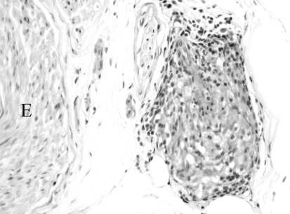
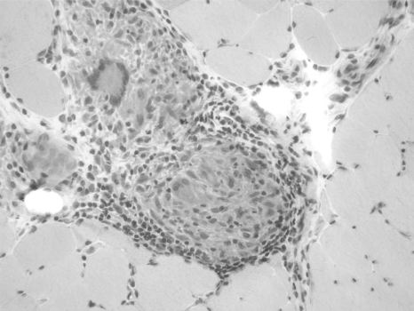
Neurosarcoidosis is an uncommon but potentially serious manifestation of sarcoidosis. While the cranial nerves are most frequently affected, neurosarcoidosis can involve other nervous system tissues including the meninges, brain parenchyma (especially the hypothalamic region), spinal cord, peripheral nerve, and muscle. Diagnosis may be particularly challenging when neurosarcoidosis occurs in isolation. Diagnostic criteria usually include histologic identification of a noncaseating granuloma, supportive laboratory or imaging tests or both, and a compatible clinical course. Treatment has not been subjected to rigorous study, but corticosteroids are typically the first line of therapy and approximately half of patients have substantial benefit. For patients who are refractory to or intolerant of corticosteroid therapy, second-line agents include azathioprine, methotrexate, cyclosporine, cyclophosphamide, mycophenolate, and even cranial irradiation. The combination of infliximab and mycophenolate mofetil is under study as well. Treatment options will likely evolve as well-designed studies are undertaken.
Keywords: Neurosarcoidosis; cranial neuropathy; meningitis; myopathy.; neuroendocrine dysfunction; peripheral neuropathy; sarcoidosis; seizures.
Figures
References
-
- Newman LS, Rose CS, Maier LA. Sarcoidosis. N. Engl. J. Med. 1997;336(17):1224–34. - PubMed
-
- Iannuzzi MC, Rybicki BA, Teirstein AS. Sarcoidosis. N. Engl. J. Med. 2007;357(21 ):2153–65. - PubMed
-
- Lower EE, Broderick JP, Brott TG, Baughman RP. Diagnosis and management of neurological sarcoidosis. Arch. Intern. Med. 1997;157(16 ):1864–8. - PubMed
-
- Stern BJ, Krumholz A, Johns C, Scott P, Nissim J. Sarcoidosis and its neurological manifestations. Arch. Neurol. 1985;42(9):909–17. - PubMed
-
- Delaney P. Neurologic manifestations in sarcoidosis: review of the literature, with a report of 23 cases. Ann. Intern. Med. 1977;87(3):336–45. - PubMed
LinkOut - more resources
Full Text Sources