Comparative Study
. 2012 Mar 6;59(10):942-53.
doi: 10.1016/j.jacc.2011.11.029.

Direct comparison of different stem cell types and subpopulations reveals superior paracrine potency and myocardial repair efficacy with cardiosphere-derived cells

Affiliations
Comparative Study

Direct comparison of different stem cell types and subpopulations reveals superior paracrine potency and myocardial repair efficacy with cardiosphere-derived cells

Tao-Sheng Li et al. J Am Coll Cardiol. .

Abstract

Objectives: The goal of this study was to conduct a direct head-to-head comparison of different stem cell types in vitro for various assays of potency and in vivo for functional myocardial repair in the same mouse model of myocardial infarction.

Background: Adult stem cells of diverse origins (e.g., bone marrow, fat, heart) and antigenic identity have been studied for repair of the damaged heart, but the relative utility of the various cell types remains unclear.

Methods: Human cardiosphere-derived cells (CDCs), bone marrow-derived mesenchymal stem cells, adipose tissue-derived mesenchymal stem cells, and bone marrow mononuclear cells were compared.

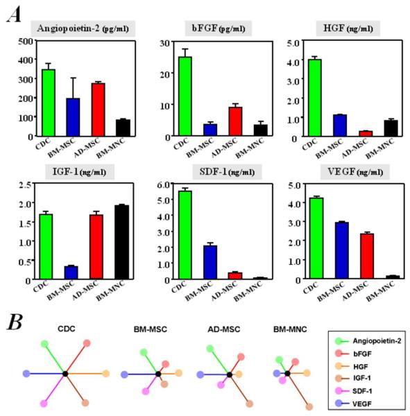
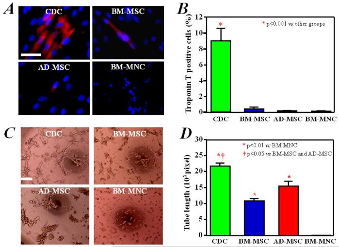
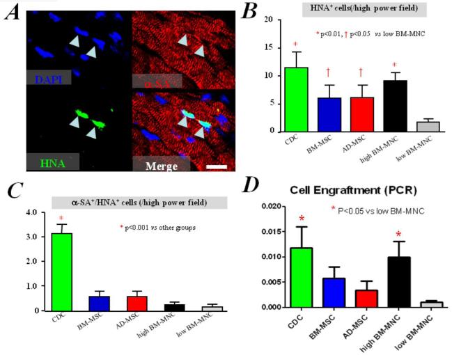
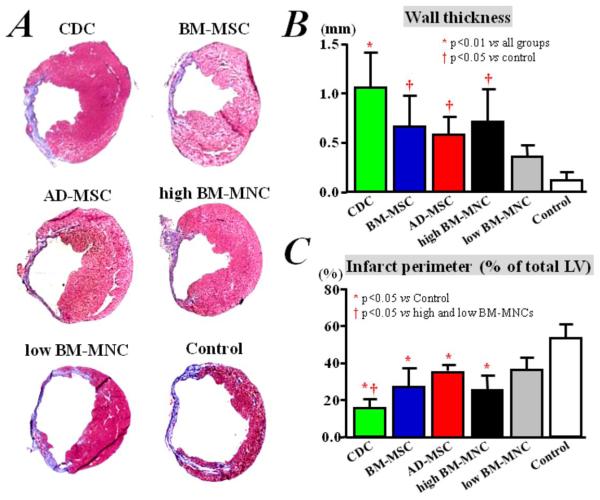
Results: CDCs revealed a distinctive phenotype with uniform expression of CD105, partial expression of c-kit and CD90, and negligible expression of hematopoietic markers. In vitro, CDCs showed the greatest myogenic differentiation potency, highest angiogenic potential, and relatively high production of various angiogenic and antiapoptotic-secreted factors. In vivo, injection of CDCs into the infarcted mouse hearts resulted in superior improvement of cardiac function, the highest cell engraftment and myogenic differentiation rates, and the least-abnormal heart morphology 3 weeks after treatment. CDC-treated hearts also exhibited the lowest number of apoptotic cells. The c-kit(+) subpopulation purified from CDCs produced lower levels of paracrine factors and inferior functional benefit when compared with unsorted CDCs. To validate the comparison of cells from various human donors, selected results were confirmed in cells of different types derived from individual rats.

Conclusions: CDCs exhibited a balanced profile of paracrine factor production and, among various comparator cell types/subpopulations, provided the greatest functional benefit in experimental myocardial infarction.


Figures

Figure 1
Figure 1. Morphology and phenotype characterization
A) Representative images of cardiosphere-derived stem cells (CDCs), bone marrow-derived mesenchymal stem cells (BM-MSCs), adipose tissue-derived mesenchymal stem cells (AD-MSCs), and bone marrow-derived mononuclear cells (BM-MNCs) after 3 days in culture under 20% O2. CDCs, BM-MSCs, and AD-MSCs demonstrate adherent growth and stromal (mesenchymal) cell-like morphology; BM-MNCs have smaller size and round shape. B) Expression profile of CD29, CD31, CD34, CD45, CD90, CD117 (c-kit), and CD133 in CDCs, BM-MSCs, AD-MSCs, and BM-MNCs. Bar = 50 μm.
Figure 2
Figure 2. Comparison of in vitro production of growth factors from cells cultured
Concentrations of angiopoietin-2, bFGF, HGF, IGF-1, SDF-1, and VEGF, measured by ELISA (n=3), are depicted (A). B) Schematic depicting the secretion of each of the six cytokines in each given cell type, as wheel-and-spoke diagrams in which the length of each spoke is proportional to the growth factor concentration in conditioned media and is normalized to that from CDCs. The symmetrical starburst pattern highlights the uniquely well-balanced paracrine profile of CDCs.
Figure 3
Figure 3. In vitro myogenic differentiation and angiogenesis assay
A) Troponin T, with distinct myocyte-like appearance, was expressed spontaneously in a fraction of CDCs cultured for 7 days. This cardiac-specific marker was rarely expressed in BM-MSCs, AD-MSCs, and BM-MNCs. B) Quantitative analysis of Troponin T expression (n=3) in CDCs (9% of the cells positive), BM-MSCs (0.4% positive) and AD-MSCs and BM-MNCs (approximately 0.1% positive). C) CDCs, BM-MSCs, and AD-MSCs produced capillary-like tube formations in extracellular matrix. BM-MNCs did not form similar structures under these conditions. D) Quantitation and comparison of tube formation capacity by the different cell types (n=3). Bars = 50 um.
Figure 4
Figure 4. Cell engraftment and in vivo myogenic differentiation
A) Immunostaining shows some human CDCs (green, HNA) expressing α-sarcomeric actin, indicating myogenic differentiation, 3 weeks after implantation into infarcted mice hearts. B) Quantitation of engraftment (HNA+ cells). C) Quantitation of cardiomyocytes differentiated from transplanted human cells (HNA+/α-SA+ cells). Bar = 20 um. D) Quantitation of engraftment by quantitative PCR (n=3 mice per group).
Figure 5
Figure 5. Cell apoptosis
A) & B) Representative images of TUNEL-positive cells in the infarcted hearts of mice 3 weeks after cell treatment with CDCs (A) and PBS (B). C) Quantitative assessment of TUNEL-positive cells in the myocardium of mice treated with different cell types and control, is shown (n=3 mice per group). Bar = 500 um.
Figure 6
Figure 6. Cardiac function
LVEF at baseline (4 hrs post-MI) did not differ among groups, indicating a similar infarct size in animals of all groups. After 3 weeks, LVEF was higher in mice implanted with CDCs, compared to controls injected with saline only. Differences between any other two groups are not significant. Data are presented as mean ± SEM.
Figure 7
Figure 7. Ventricular remodeling
A) Representative images of Masson’s staining of infarcted mice hearts, after implantation of different types of human cells or saline injection only. Quantitative analyses (n=3 mice per group) of LV wall thickness (B) and infarct perimeter (C) show that remodeling was attenuated more efficiently by CDC implantation, compared with BM-MSCs, AD-MSCs, and BM-MNC treatment, although implantation of BM-MSCs, AD-MSCs, and BM-MNCs resulted in less remodeling compared to control treatment with saline injection only.
Figure 8
Figure 8. Comparison of purified c-kit+ stem cells and unsorted CDCs
A) 3 weeks after infarction, LVEF was higher in mice that received unsorted CDCs than those with c-kit+ cells purified from the same CDCs, although these purified c-kit+ stem cells also improved cardiac function when compared to controls injected with saline only. B) Although the same number of cells was used for culture, the purified c-kit+ stem cells released less VEGF, SDF, IGF-1, and HGF than the unsorted CDCs.

References

    1. Beltrami AP, Barlucchi L, Torella D, et al. Adult cardiac stem cells are multipotent and support myocardial regeneration. Cell. 2003;114:763–76. - PubMed
    1. Oh H, Bradfute SB, Gallardo TD, et al. Cardiac progenitor cells from adult myocardium: homing, differentiation, and fusion after infarction. Proc Natl Acad Sci USA. 2003;100:12313–8. - PMC - PubMed
    1. Matsuura K, Nagai T, Nishigaki N, et al. Adult cardiac Sca-1-positive cells differentiate into beating cardiomyocytes. J Biol Chem. 2004;279:11384–91. - PubMed
    1. Pfister O, Mouquet F, Jain M, et al. CD31-but Not CD31+ cardiac side population cells exhibit functional cardiomyogenic differentiation. Circ Res. 2005;97:52–61. - PubMed
    1. Laugwitz KL, Moretti A, Lam J, et al. Postnatal isl1+ cardioblasts enter fully differentiated cardiomyocyte lineages. Nature. 2005;433:647–53. - PMC - PubMed

Publication types

MeSH terms