

Evaluation of disease and viral biomarkers as triggers for therapeutic intervention in respiratory mousepox - an animal model of smallpox
- PMID: 22381921
- PMCID: PMC3722602
- DOI: 10.1016/j.antiviral.2012.02.005
Evaluation of disease and viral biomarkers as triggers for therapeutic intervention in respiratory mousepox - an animal model of smallpox
Abstract
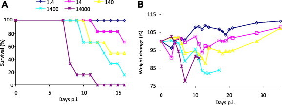
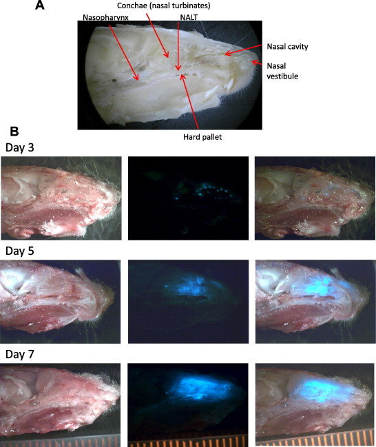
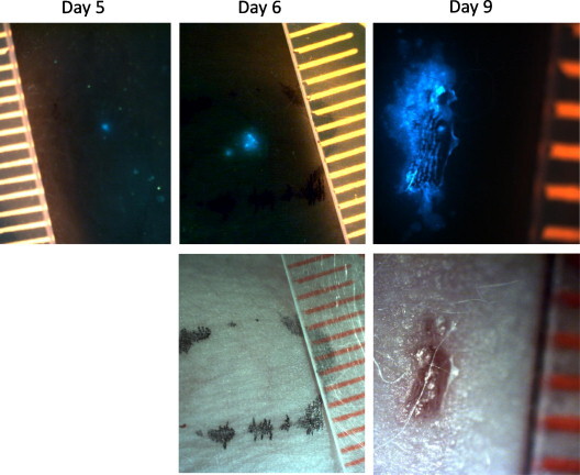
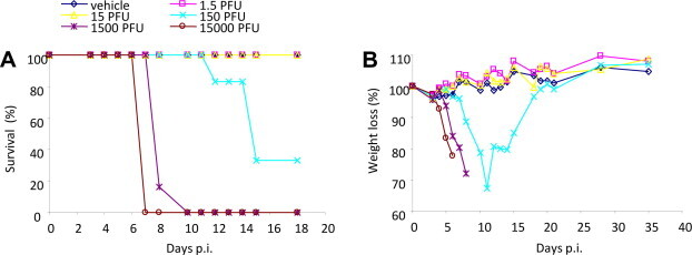
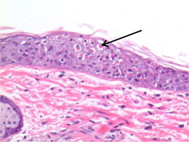
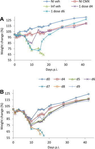
The human population is currently faced with the potential use of natural or recombinant variola and monkeypox viruses as biological weapons. Furthermore, the emergence of human monkeypox in Africa and its expanding environs poses a significant natural threat. Such occurrences would require therapeutic and prophylactic intervention with antivirals to minimize morbidity and mortality of exposed populations. Two orally-bioavailable antivirals are currently in clinical trials; namely CMX001, an ether-lipid analog of cidofovir with activity at the DNA replication stage and ST-246, a novel viral egress inhibitor. Both of these drugs have previously been evaluated in the ectromelia/mousepox system; however, the trigger for intervention was not linked to a disease biomarker or a specific marker of virus replication. In this study we used lethal, intranasal, ectromelia virus infections of C57BL/6 and hairless SKH1 mice to model human disease and evaluate exanthematous rash (rash) as an indicator to initiate antiviral treatment. We show that significant protection can be provided to C57BL/6 mice by CMX001 or ST-246 when therapy is initiated on day 6 post infection or earlier. We also show that significant protection can be provided to SKH1 mice treated with CMX001 at day 3 post infection or earlier, but this is four or more days before detection of rash (ST-246 not tested). Although in this model rash could not be used as a treatment trigger, viral DNA was detected in blood by day 4 post infection and in the oropharyngeal secretions (saliva) by day 2-3 post infection - thus providing robust and specific markers of virus replication for therapy initiation. These findings are discussed in the context of current respiratory challenge animal models in use for the evaluation of poxvirus antivirals.
Copyright © 2012 Elsevier B.V. All rights reserved.
Figures
Similar articles
-
Buccal viral DNA as a trigger for brincidofovir therapy in the mousepox model of smallpox.Antiviral Res. 2017 Mar;139:112-116. doi: 10.1016/j.antiviral.2016.12.015. Epub 2016 Dec 27. Antiviral Res. 2017. PMID: 28039021 Free PMC article.
-
Efficacy of therapeutic intervention with an oral ether-lipid analogue of cidofovir (CMX001) in a lethal mousepox model.Antiviral Res. 2008 Jan;77(1):39-49. doi: 10.1016/j.antiviral.2007.08.003. Epub 2007 Sep 4. Antiviral Res. 2008. PMID: 17904231 Free PMC article.
-
Using biomarkers to stage disease progression in a lethal mousepox model treated with CMX001.Antivir Ther. 2008;13(7):863-73. Antivir Ther. 2008. PMID: 19043920 Free PMC article.
-
Efficacy of CMX001 as a post exposure antiviral in New Zealand White rabbits infected with rabbitpox virus, a model for orthopoxvirus infections of humans.Viruses. 2011 Jan;3(1):47-62. doi: 10.3390/v3010047. Viruses. 2011. PMID: 21373379 Free PMC article. Review.
-
Efficacy of CMX001 as a prophylactic and presymptomatic antiviral agent in New Zealand white rabbits infected with rabbitpox virus, a model for orthopoxvirus infections of humans.Viruses. 2011 Feb;3(2):63-82. doi: 10.3390/v3020063. Viruses. 2011. PMID: 21369346 Free PMC article. Review.
Cited by
-
GETTING AHEAD OF MONKEYPOX: Learning from the COVID-19 pandemic experience to prevent the potentially new monkeypox pandemic.J Med Virol. 2023 Jan;95(1):e28146. doi: 10.1002/jmv.28146. Epub 2022 Sep 26. J Med Virol. 2023. PMID: 36129007 Free PMC article.
-
Orthopoxvirus inhibitors that are active in animal models: an update from 2008 to 2012.Future Virol. 2013 Sep;8(9):891-901. doi: 10.2217/fvl.13.76. Future Virol. 2013. PMID: 24563659 Free PMC article.
-
Clinical and Translational Pharmacology Considerations for Anti-infectives Approved Under the FDA Animal Rule.Clin Pharmacokinet. 2023 Jul;62(7):943-953. doi: 10.1007/s40262-023-01267-x. Epub 2023 Jun 16. Clin Pharmacokinet. 2023. PMID: 37326917 Free PMC article. Review.
-
A review of experimental and natural infections of animals with monkeypox virus between 1958 and 2012.Future Virol. 2013 Feb 1;8(2):129-157. doi: 10.2217/fvl.12.130. Future Virol. 2013. PMID: 23626656 Free PMC article.
-
Monkeypox: epidemiology, pathogenesis, treatment and prevention.Signal Transduct Target Ther. 2022 Nov 2;7(1):373. doi: 10.1038/s41392-022-01215-4. Signal Transduct Target Ther. 2022. PMID: 36319633 Free PMC article. Review.
References
-
- Bilder L., Machtei E.E., Shenhar Y., Kra-Oz Z., Basis F. Salivary detection of H1N1 virus: a clinical feasibility investigation. J. Dent. Res. 2011;90:1136–1139. - PubMed
-
- Boppana S.B., Ross S.A., Shimamura M., Palmer A.L., Ahmed A., Michaels M.G., Sanchez P.J., Bernstein D.I., Tolan R.W., Jr., Novak Z., Chowdhury N., Britt W.J., Fowler K.B. Saliva polymerase-chain-reaction assay for cytomegalovirus screening in newborns. N. Engl. J. Med. 2011;364:2111–2118. - PMC - PubMed
Publication types
MeSH terms
Substances
Grants and funding
LinkOut - more resources
Full Text Sources
Medical
