

Phytobezoar in Meckel's diverticulum: A rare cause of small bowel obstruction
- PMID: 22382033
- PMCID: PMC3316763
- DOI: 10.1016/j.ijscr.2012.01.006
Phytobezoar in Meckel's diverticulum: A rare cause of small bowel obstruction
Abstract
Introduction: Meckel's diverticulum (MD) is the prevailing anomaly of the gastrointestinal tract, found in about 2% of the population; it rarely gives rise to symptoms and its discovery is usually accidental. Phytobezoar is a concretion of poorly digested fruit and vegetable fibres that is found in the alimentary tract and rarely can be the cause of small intestinal obstruction. Herein we report a rare case of intestinal obstruction due to phytobezoar formation into a MD.
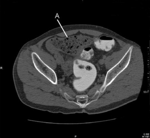
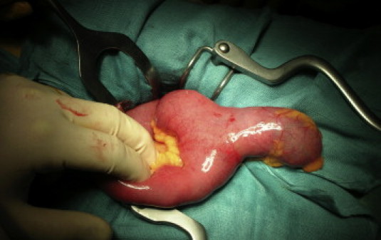
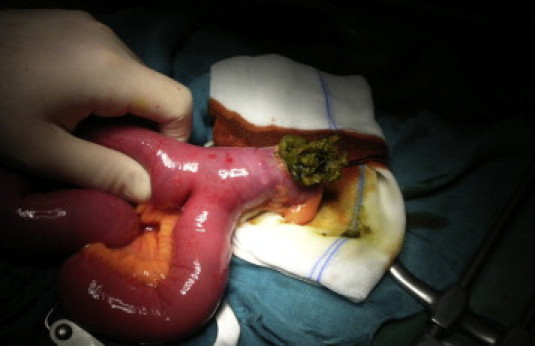
Presentation of case: A 50 year-old patient, was admitted to author's institution with an history of abdominal pain, nausea and multiples episodes of vomiting. Plain X-ray showed dilated small-bowel loops. Computed tomography (CT) revealed jejunal loops with air-fluid levels. The patient underwent explorative laparotomy where we found a giant Meckel's diverticulum, filled by a phytobezoar that caused small bowel compression. We performed a segmental ileal, resection, containing the MD. The histological exam confirmed Meckel's diverticulum.
Discussion: Bowel obstruction due to a phytobezoar in a Meckel's diverticulum is rare: only 7 cases have been reported in literature. MD complications are rare and phytobezoar is one of them with only few cases described in literature.
Conclusion: The conventional x rays studies were inconclusive whereas abdominal contrast enhanced CT led to a definitive diagnosis. Explorative laparotomy or laparoscopy is mandatory in these cases.
Copyright © 2012 Surgical Associates Ltd. Published by Elsevier Ltd. All rights reserved.
Figures
References
-
- Levy A.D., Hobbs C.M., From the archives of the AFIP Meckel's diverticulum: radiologic features with pathologic correlation. Radiographics. 2004;24:565–587. - PubMed
-
- Soltero M.J., Bill A.H. The natural history of Meckel's Diverticulum and its relation to incidental removal. A study of 202 cases of diseased Meckel's Diverticulum found in King County, Washington, over a fifteen year period. Am J Surg. 1976;132:168–173. - PubMed
-
- Vane D.W., West K.W., Grosfeld J.L. Vitelline duct anomalies. Experience with 217 childhood cases. Arch Surg. 1987;122:542–547. - PubMed
-
- Ousadden A., Mazaz K., Mellouki I., Taleb K.A. Gastric trichobezoar: one case report. Ann Chir. 2004;129:237–240. - PubMed
-
- Tulin Y., Sedat Y., OPzlen B., Levent O., Turgut N. Small bowel obstruction due to phytobezoar: CT diagnosis. Eur Radiol. 2002;12:2659–2661. - PubMed
LinkOut - more resources
Full Text Sources
Research Materials
