Review
. 2012 Nov;135(Pt 11):3206-26.
doi: 10.1093/brain/aws023. Epub 2012 Mar 1.

Cerebral causes and consequences of parkinsonian resting tremor: a tale of two circuits?

Affiliations
Review

Cerebral causes and consequences of parkinsonian resting tremor: a tale of two circuits?

Rick C Helmich et al. Brain. 2012 Nov.

Abstract

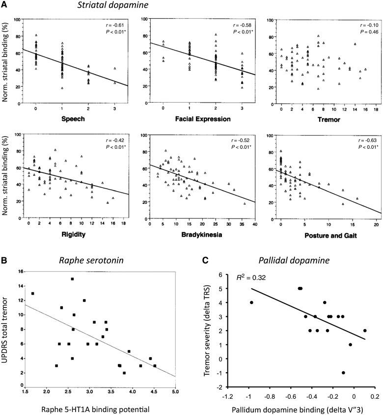
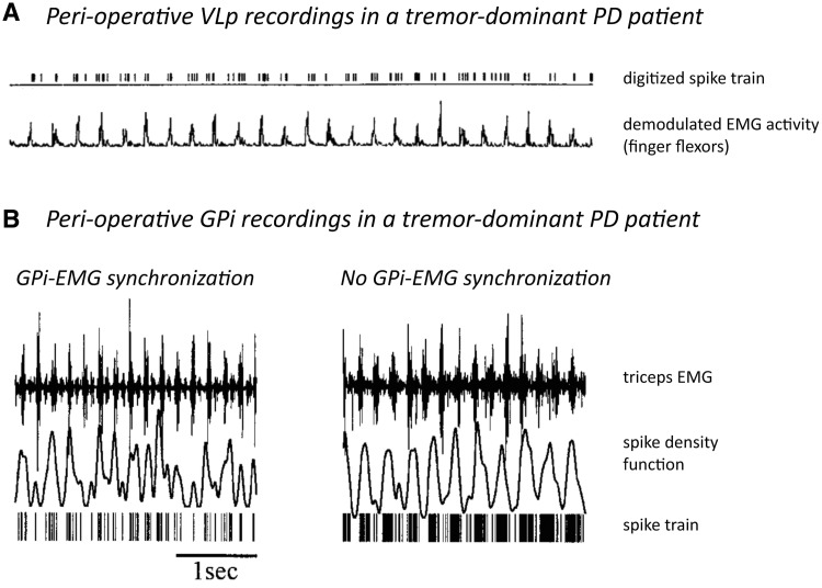
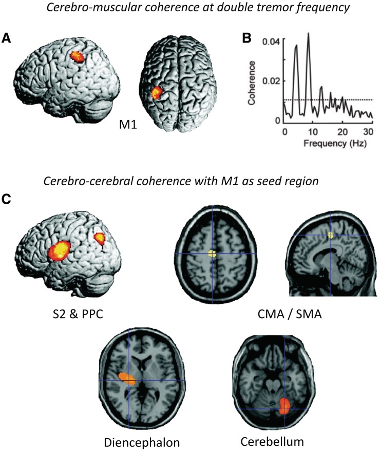
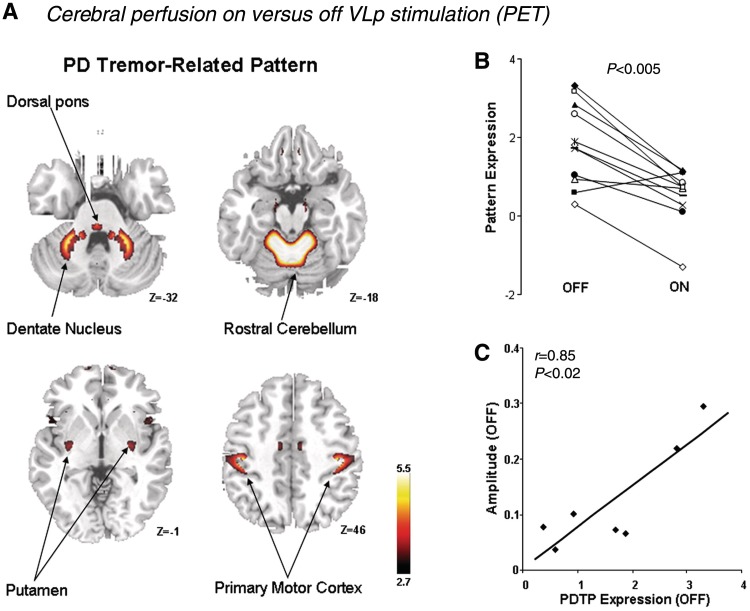
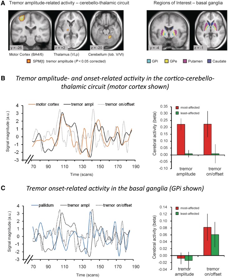
Tremor in Parkinson's disease has several mysterious features. Clinically, tremor is seen in only three out of four patients with Parkinson's disease, and tremor-dominant patients generally follow a more benign disease course than non-tremor patients. Pathophysiologically, tremor is linked to altered activity in not one, but two distinct circuits: the basal ganglia, which are primarily affected by dopamine depletion in Parkinson's disease, and the cerebello-thalamo-cortical circuit, which is also involved in many other tremors. The purpose of this review is to integrate these clinical and pathophysiological features of tremor in Parkinson's disease. We first describe clinical and pathological differences between tremor-dominant and non-tremor Parkinson's disease subtypes, and then summarize recent studies on the pathophysiology of tremor. We also discuss a newly proposed 'dimmer-switch model' that explains tremor as resulting from the combined actions of two circuits: the basal ganglia that trigger tremor episodes and the cerebello-thalamo-cortical circuit that produces the tremor. Finally, we address several important open questions: why resting tremor stops during voluntary movements, why it has a variable response to dopaminergic treatment, why it indicates a benign Parkinson's disease subtype and why its expression decreases with disease progression.


Figures

Figure 1
Figure 1
Neurochemical correlates of Parkinson's disease tremor. (A) Correlation between age-normalized striatal [123] I beta-CIT binding and UPDRS motor subscores for speech, facial expression, tremor (rest and action tremor), rigidity, bradykinesia, and posture and gait (n = 59). Reprinted from Pirker (2003), with permission from John Wiley and Sons. (B) Correlation between 11C-WAY 100635 PET in the raphe and total UPDRS tremor score (r = −0529; P < 0.01; n = 23). Reprinted from Doder et al. (2003), with permission from Wolters Kluwer. (C) Correlation between pallidal [I-123] FP-CIT binding and resting tremor severity [tremor rating scale (TRS); r = −0.57; P = 0.023], using within-patient difference scores (between most-affected and least-affected sides). This procedure controls for non-specific differences between patients. Reprinted from Helmich et al. (2011b), with permission from John Wiley and Sons. These data show that tremor severity is correlated with dopamine depletion in the pallidum (C), but not the striatum (A), and also with serotonin depletion in the raphe (B).
Figure 2
Figure 2
Neuronal correlates of Parkinson's disease tremor. (A) Simultaneous recording of thalamic posterior VL (VLp) single-unit activity and peripheral EMG during tremor in a parkinsonian patient. These data show continuous synchronization between internal globus pallidus activity and peripheral EMG. Reprinted from Lenz et al. (1988), with permission from the Society for Neuroscience. (B) Simultaneous recording of internal globus pallidus (GPi) multi-unit activity and peripheral EMG during tremor in a patient with Parkinson's disease (PD). The two plots illustrate the raw signals of two epochs of data sampled 5 min apart. Note that in the left trace the peaks in the spike density function coincide with the EMG bursts, whereas in the right trace the oscillations in the spike density function occur at a lower frequency than the EMG. These data show that synchronization between neuronal activity in internal globus pallidus and peripheral EMG is transient in nature. Reprinted from Hurtado et al. (1999). Copyright (2011) National Academy of Sciences, USA.
Figure 3
Figure 3
Oscillatory correlates of Parkinson's disease tremor. This figure shows statistical parametric (SPM) maps of spatially normalized cerebro-muscular and cerebro-cerebral coherence of four patients with right-sided rest tremor. Cerebral activity was measured with magnetoencephalography and muscular activity with EMG. (A) Cerebro-muscular coherence at double tremor frequency is located in the contralateral primary motor cortex (M1). (B) This plot shows the coherence between M1 activity and the tremor EMG for one patient with Parkinson's disease. The dashed line indicates the 99% confidence level. (C) Cerebro-cerebral coherence was computed with the reference region in M1 and averaged for all four patients. Areas of consistent coupling with M1 were found in the secondary somatosensory cortex (S2), posterior parietal cortex (PPC), cingulate motor area (CMA)/supplementary motor area (SMA), contralateral diencephalon and ipsilateral cerebellum. Due to the poor coverage by and the large distance to the magnetoencephalography sensors, localization in the latter two areas is not as precise as at the cortical level. These data show a cerebello-thalamo-cortical circuit coupled with tremor on a cycle-by-cycle basis. Reprinted from Timmermann et al. (2003), by permission of Oxford University Press.
Figure 4
Figure 4
Metabolic correlates of Parkinson's disease tremor. (A) Spatial covariance pattern identified by ordinal trends canonical variate analysis of FDG PET data from 11 hemispheres of nine patients with tremor-dominant Parkinson's disease (PD) scanned on and off posterior VL stimulation (labelled ventral intermediate nucleus in the original manuscript). Posterior VL stimulation improved tremor severity and reduced metabolic activity in the primary motor cortex, anterior cerebellum/dorsal pons, and the caudate/putamen. (B) The expression of this Parkinson's disease tremor-related metabolic pattern (PDTP) was reduced by posterior VL stimulation in 10 of the 11 treated hemispheres. (C) Baseline PDTP expression (i.e. off-stimulation pattern scores) correlated (r = 0.85, P < 0.02) with tremor amplitude, measured with concurrent accelerometry. These data show that metabolic activity in both the cortico-cerebellar circuit and the basal ganglia is related to tremor severity. Reprinted from Mure et al. (2011), with permission from Elsevier.
Figure 5
Figure 5
Tremor amplitude- and onset-related cerebral activity in Parkinson's disease. (A) Left: Cerebral regions where activity co-fluctuated with tremor amplitude (19 tremor-dominant patients, P < 0.05 whole-brain corrected). Activity was localized to the motor cortex, thalamus (posterior VL; VLp) and cerebellum (left side = side contralateral to tremor). Right: Regions of interest in the basal ganglia are shown. (B) In the cerebello-thalamo-cortical circuit, we found two separate effects: (i) cerebral activity related to tremor amplitude and (ii) cerebral activity related to changes in tremor amplitude (tremor on/offset). Left: These two tremor-related effects are illustrated for the motor cortex of one patient. Right: These two tremor-related effects are shown for the motor cortex across the whole group (19 tremor-dominant patients), separately for the most- and least-affected hemisphere. Similar effects were found in the posterior VL and cerebellum (not shown). (C) In the basal ganglia, we found cerebral activity related to changes in tremor amplitude (tremor on/offset), but not cerebral activity related to tremor amplitude. Left: This effect is illustrated for the internal globus pallidus of one patient. Right: This effect is shown for the internal globus pallidus (GPi) across the whole group (19 tremor-dominant patients), separately for the most- and least-affected hemisphere. Similar effects were found for the putamen, but not for the caudate (not shown). The line graphs in B and C show three relevant time courses: (i) brain activity (motor cortex in orange, internal globus pallidus in blue); (ii) tremor amplitude of the contralateral hand (in black; EMG regressor convolved with the haemodynamic response function); and (iii) tremor on/offset (in dotted grey, first temporal derivative of the tremor amplitude regressor, convolved with the haemodynamic response function). These data suggest distinct contributions of two circuits to tremor: the cerebello-thalamo-cortical circuit controls tremor amplitude, and the striato-pallidal circuit produces changes in tremor amplitude. Reprinted from Helmich et al. (2011b), with permission from John Wiley and Sons. GPe = external globus pallidus.
Figure 6
Figure 6
The dimmer-switch model of parkinsonian resting tremor. In tremor-dominant Parkinson's disease, dopaminergic cell death in the retrorubral area A8 causes dopamine depletion in the pallidum (in red). Pallidal dopamine depletion leads to emergence of pathological activity in the striato-pallidal circuit, which triggers activity in the cerebello-thalamo-cortical circuit (in blue) through the primary motor cortex (red line between pallidum and primary motor cortex). Thus, the striato-pallidal circuit triggers tremor episodes (analogues to a light switch), while the cerebello-thalamo-cortical circuit produces the tremor and controls its amplitude (analogous to a light dimmer). This model is based on Helmich et al. (2011b). VLp = posterior VL.
Figure 7
Figure 7
A beat of tremor preceding movement in Parkinson's disease. Fast flexion patterns in patients with Parkinson's disease. In many patients with Parkinson's disease (17 arms of 15 patients), although resting tremor was not continuously present, a single ‘beat of tremor’ occasionally occurred before the pattern that moved the limb. This is illustrated for one patient. Adapted from Hallett et al. (1977) with permission from BMJ Publishing Group Ltd.

Similar articles

Cited by

References

    1. Aarsland D, Andersen K, Larsen JP, Lolk A, Kragh-Sorensen P. Prevalence and characteristics of dementia in Parkinson disease: an 8-year prospective study. Arch Neurol. 2003;60:387–92. - PubMed
    1. Abdo WF, van de Warrenburg BP, Burn DJ, Quinn NP, Bloem BR. The clinical approach to movement disorders. Nat Rev Neurol. 2010;6:29–37. - PubMed
    1. Albin RL, Young AB, Penney JB. The functional anatomy of basal ganglia disorders. Trends Neurosci. 1989;12:366–75. - PubMed
    1. Albo Z, Di Prisco GV, Chen YH, Rangarajan G, Truccolo W, Feng JF, Vertes RP, Ding MZ. Is partial coherence a viable technique for identifying generators of neural oscillations? Biol Cybernet. 2004;90:318–26. - PubMed
    1. Alves G, Larsen JP, Emre M, Wentzel-Larsen T, Aarsland D. Changes in motor subtype and risk for incident dementia in Parkinson's disease. Mov Disord. 2006;21:1123–30. - PubMed

Publication types

MeSH terms