Review
. 2012 Mar;5(2):155-64.
doi: 10.1242/dmm.009019.

Biogenesis of iron-sulfur clusters in mammalian cells: new insights and relevance to human disease

Affiliations
Review

Biogenesis of iron-sulfur clusters in mammalian cells: new insights and relevance to human disease

Tracey A Rouault. Dis Model Mech. 2012 Mar.

Abstract

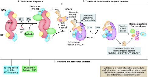
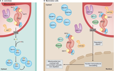
Iron-sulfur (Fe-S) clusters are ubiquitous cofactors composed of iron and inorganic sulfur. They are required for the function of proteins involved in a wide range of activities, including electron transport in respiratory chain complexes, regulatory sensing, photosynthesis and DNA repair. The proteins involved in the biogenesis of Fe-S clusters are evolutionarily conserved from bacteria to humans, and many insights into the process of Fe-S cluster biogenesis have come from studies of model organisms, including bacteria, fungi and plants. It is now clear that several rare and seemingly dissimilar human diseases are attributable to defects in the basic process of Fe-S cluster biogenesis. Although these diseases -which include Friedreich's ataxia (FRDA), ISCU myopathy, a rare form of sideroblastic anemia, an encephalomyopathy caused by dysfunction of respiratory chain complex I and multiple mitochondrial dysfunctions syndrome - affect different tissues, a feature common to many of them is that mitochondrial iron overload develops as a secondary consequence of a defect in Fe-S cluster biogenesis. This Commentary outlines the basic steps of Fe-S cluster biogenesis as they have been defined in model organisms. In addition, it draws attention to refinements of the process that might be specific to the subcellular compartmentalization of Fe-S cluster biogenesis proteins in some eukaryotes, including mammals. Finally, it outlines several important unresolved questions in the field that, once addressed, should offer important clues into how mitochondrial iron homeostasis is regulated, and how dysfunction in Fe-S cluster biogenesis can contribute to disease.


Figures

Fig. 1.
Fig. 1.
A general scheme for biogenesis of Fe-S clusters in mammalian cells. (A) NFS1 is a cysteine desulfurase that forms a dimer to which monomers of the primary scaffold protein ISCU bind near the top and bottom of the complex. In eukaryotes, ISD11 is an obligate binding partner for NFS1. NFS1 also binds the cofactor pyridoxal phosphate (not shown). Structural and biochemical studies suggest that frataxin forms part of the initial Fe-S cluster biogenesis complex, potentially occupying a pocket between NFS1 and ISCU. NFS1 donates inorganic sulfur, and cysteines from ISCU provide the sulfur ligands that directly bind iron in the nascent Fe-S cluster. A highly reduced protein such as ferredoxin probably provides needed electrons. (B) Once the Fe-S cluster is assembled, it must be transferred to recipient proteins. Work in bacteria and yeast model systems suggests that a dedicated chaperone–co-chaperone pair of proteins participates in cluster transfer from the primary scaffold, ISCU, to recipient Fe-S proteins. The co-chaperone is known to be HSC20 (a DNAJ protein), whereas the chaperone is an HSP70 homolog that has not yet been clearly identified in mammalian cells. The role of a putative HSC70 protein is proposed here. HSC20 binds ISCU, and the HSC20-ISCU complex probably then binds to its HSC70 partner through two different binding sites: HSC20 contacts the N-terminus of HSC70 and its binding partner, ISCU, binds to the C-terminal substrate-binding domain region of HSC70. The J domain region of HSC20 contains three residues [His (H) Pro (P) and Asp (D); HPD] that activate the ATPase activity of HSC70. Upon activation, a conformational change is proposed to occur in the substrate-binding domain of HSC70 that affects bound ISCU, resulting in extrusion of a peptide containing the residues LPPVK from the ISCU globular protein. The LPPVK peptide then binds to a groove in the substrate-binding domain of HSC70, which consolidates or perhaps further enhances the conformational change in ISCU, which might convert it to a conformation that facilitates donation of its cluster to recipient proteins. In this model, HSC20 helps protect the vulnerable Fe-S cluster bound to ISCU as it dissociates from the multimeric assembly complex, and HSC20 then escorts ISCU to form a trimeric complex with HSC70. The consumption of ATP probably provides a powerful impetus to drive conformational changes of ISCU and the substrate-binding domain of HSC70; these changes might facilitate release of the Fe-S cluster from ISCU. By capturing the energy released by ATP hydrolysis and coupling it to conformational changes, the chaperone–co-chaperone pair help the Fe-S cluster to reach its target proteins. Target proteins could include some direct targets, or proteins such as NFU1, BOLA3, NUBPL or GLRX5 that might function as intermediary scaffolds that then donate Fe-S clusters to specific subsets of recipient proteins. (C) Mutations in proteins acting at different points in the biogenesis pathway cause diseases with markedly different phenotypes (described in Table 1).
Fig. 2.
Fig. 2.
Proposed differences in the localization of Fe-S cluster biogenesis proteins in S. cerevisiae versus mammalian cells. (A) Yeast mitochondria contain the basic Fe-S cluster biogenesis proteins, whereas cytosolic Fe-S cluster biogenesis has been proposed to depend on the export of a sulfur compound from mitochondria through the transporter Atm1 (ABCB7 in human cells). Further biogenesis in the cytoplasm depends on a distinct set of proteins, including Tah18, Dre2, Nbp35, Cfd1, Nar1 and Cia1, which are collectively referred to as CIA (cytosolic iron-sulfur assembly) proteins. (B) By contrast, in mammalian cells, it seems that most of the basic Fe-S cluster biogenesis proteins are expressed in mitochondria as well as in the cytosolic and/or nuclear compartment (nuclear localization of these proteins not represented in the figure). In addition, there are human counterparts to the proteins implicated in cytosolic Fe-S cluster biogenesis in yeast, including NDOR1 (homolog of Tah18), CIAPIN1, (homolog of Dre2), NUBP1 and NUBP2 (homologs of Nbp35 and Cfd1, respectively), and NARFL and CIAO1 (homologs of Nar1 and Cia1, respectively). To explain mitochondrial iron overload (which occurs as a consequence of disrupted Fe-S cluster biogenesis), it is possible that mitochondria depend on Fe-S cluster biogenesis to synthesize a molecule that functions as a regulatory signal. In the absence of this signal (i.e. when Fe-S cluster biogenesis is impaired), remodeling of nuclear transcription leads to mitochondrial iron overload, as the cell attempts to compensate for possible mitochondrial iron deficiency. Because mitochondrial iron overload probably contributes to disease pathogenesis in FRDA, ISCU myopathy and possibly several other diseases, elucidation of the details of this pathway might pave the way to new therapeutic interventions.

Similar articles

Cited by

References

    1. Adam A. C., Bornhovd C., Prokisch H., Neupert W., Hell K. (2006). The Nfs1 interacting protein Isd11 has an essential role in Fe/S cluster biogenesis in mitochondria. EMBO J. 25, 174–183 - PMC - PubMed
    1. Allikmets R., Raskind W. H., Hutchinson A., Schueck N. D., Dean M., Koeller D. M. (1999). Mutation of a putative mitochondrial iron transporter gene (ABC7) in X-linked sideroblastic anemia and ataxia (XLSA/A). Hum. Mol. Genet. 8, 743–749 - PubMed
    1. Babcock M., de Silva D., Oaks R., Davis-Kaplan S., Jiralerspong S., Montermini L., Pandolfo M., Kaplan J. (1997). Regulation of mitochondrial iron accumulation by Yfh1p, a putative homolog of frataxin. Science 276, 1709–1712 - PubMed
    1. Bandyopadhyay S., Chandramouli K., Johnson M. K. (2008). Iron-sulfur cluster biosynthesis. Biochem. Soc. Trans. 36, 1112–1119 - PMC - PubMed
    1. Bedekovics T., Li H., Gajdos G. B., Isaya G. (2011). Leucine biosynthesis regulates cytoplasmic iron-sulfur enzyme biogenesis in an Atm1-independent manner. J. Biol. Chem. 286, 40878–40888 - PMC - PubMed

Publication types

MeSH terms

Substances