. 2012 May;33(5):976-85.
doi: 10.1093/carcin/bgs114. Epub 2012 Mar 1.

MiRNA-335 suppresses neuroblastoma cell invasiveness by direct targeting of multiple genes from the non-canonical TGF-β signalling pathway

Affiliations

MiRNA-335 suppresses neuroblastoma cell invasiveness by direct targeting of multiple genes from the non-canonical TGF-β signalling pathway

Jennifer Lynch et al. Carcinogenesis. 2012 May.

Abstract

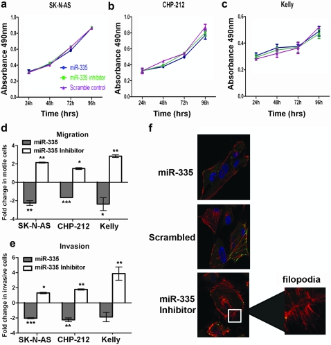
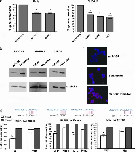
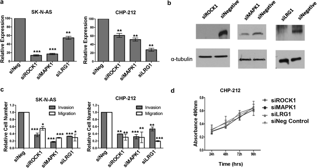
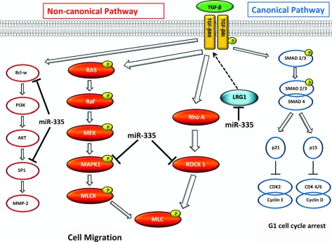
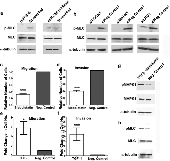
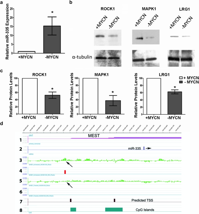
Transforming growth factor-β (TGF-β) signaling regulates many diverse cellular activities through both canonical (SMAD-dependent) and non-canonical branches, which includes the mitogen-activated protein kinase (MAPK), Rho-like guanosine triphosphatase and phosphatidylinositol-3-kinase/AKT pathways. Here, we demonstrate that miR-335 directly targets and downregulates genes in the TGF-β non-canonical pathways, including the Rho-associated coiled-coil containing protein (ROCK1) and MAPK1, resulting in reduced phosphorylation of downstream pathway members. Specifically, inhibition of ROCK1 and MAPK1 reduces phosphorylation levels of the motor protein myosin light chain (MLC) leading to a significant inhibition of the invasive and migratory potential of neuroblastoma cells. Additionally, miR-335 targets the leucine-rich alpha-2-glycoprotein 1 (LRG1) messenger RNA, which similarly results in a significant reduction in the phosphorylation status of MLC and a decrease in neuroblastoma cell migration and invasion. Thus, we link LRG1 to the migratory machinery of the cell, altering its activity presumably by exerting its effect within the non-canonical TGF-β pathway. Moreover, we demonstrate that the MYCN transcription factor, whose coding sequence is highly amplified in a particularly clinically aggressive neuroblastoma tumor subtype, directly binds to a region immediately upstream of the miR-335 transcriptional start site, resulting in transcriptional repression. We conclude that MYCN contributes to neuroblastoma cell migration and invasion, by directly downregulating miR-335, resulting in the upregulation of the TGF-β signaling pathway members ROCK1, MAPK1 and putative member LRG1, which positively promote this process. Our results provide novel insight into the direct regulation of TGF-β non-canonical signaling by miR-335, which in turn is downregulated by MYCN.


Figures

Fig. 1.
Fig. 1.
miR-335 regulates the migratory and invasive potential of neuroblastoma cell lines and does not affect cell viability. (3-(4,5-dimethylthiazol-2-yl)-5-(3-carboxymethoxyphenyl)-2-(4-sulfophenyl)-2H-tetrazolium) cell viability assays following transfection of SK-N-AS (a), CHP-212 (b) and Kelly (c) cells with mature miR-335 mimics, miR-335 inhibitor or scrambled negative control. (d and e) Analysis of the ability of SK-N-AS, CHP-212 and Kelly cells to migrate across a non-matrigel membrane (d) and to invade across a matrigel membrane (e) following transfection with mature miR-335 mimics, miR-335 inhibitor or scrambled control. Asterisks indicate statistical significance obtained by unpaired Students’s t-test. *P < 0.05, **P < 0.005, ***P < 0.0005. (f) Staining of F-actin and focal adhesions in SK-N-AS cells with fluorescence-labeled phalloidin (red) and vinculin (green), respectively. Cell nuclei are stained with 4′,6-diamidino-2-phenylindole (blue). An enlarged image of the filopodia in miR-335 inhibitor-transfected cells is indicated by the white box.
Fig. 2.
Fig. 2.
miR-335 target gene identification and validation. (a) Reverse transcription–qPCR expression analysis of ROCK1, MAPK1 and LRG1 in Kelly and CHP-212 cells following transfection with mature miR-335 mimics. LRG1 expression could not be detected in Kelly cells. (b) Western blot analysis revealed reduced expression of ROCK1, MAPK1 and LRG1 in CHP-212 cells transfected with miR-335. (c) Immunofluorescent staining of Kelly cells facilitated visualization of reduced ROCK1 protein expression in miR-335-transfected cells and increased expression of ROCK1 in miR-335 inhibitor-transfected cells. ROCK1 (red), 4′,6-diamidino-2-phenylindole (blue). (d) Luciferase reporter assays confirmed direct targeting of ROCK1, MAPK1 and LRG1 3′UTR regions by miR-335. A significant reduction in luciferase activity was measured for each of the wild-type miR-335 binding sites for each of the three genes compared with the mutated binding sites, normalized against scrambled control. The miRNA:mRNA base pairing for each gene is also illustrated. *P < 0.05, **P < 0.005.
Fig. 3.
Fig. 3.
siRNA-mediated functional effects of ROCK1, MAPK1 and LRG1. (a) Reverse transcription–qPCR analysis demonstrated significant reductions in the expression levels of ROCK1, MAPK1 and LRG1 in SK-N-AS and CHP-212 cells 48 h post-transfection. (b) Western blot analysis also revealed significant reduction in protein levels of ROCK1, MAPK1 and LRG1 72 h post-transfection of CHP-212 cells. (c) SK-N-AS and CHP-212 cells demonstrated significant reductions in migration and invasion following knockdown of ROCK1, MAPK1 and LRG1. (d) Knockdown of ROCK1, MAPK1 and LRG1 did not alter the rate of proliferation of CHP-212 cells as compared with siNegative control transfected cells. *P < 0.05, **P < 0.005, ***P < 0.0005.
Fig. 4.
Fig. 4.
Schematic illustration of TGF-β signaling pathway members and potential pathway member LRG1. TGF-β pathway comprises the canonical and non-canonical signaling pathways. miR-335 regulates cell migration and invasion through targeting of the three branches of the non-canonical TGF-β pathway. In this study, miR-335 has been demonstrated to directly target ROCK1, MAPK1 and putative TGF-β member LRG1. miR-335 inhibition of ROCK1 prevents phosphorylation of MLC thereby reducing actomyosin assembly and contraction and ultimately the motile potential of the cells. Similarly, inhibition of MAPK1 signaling also prevents phosphorylation of MLC.
Fig. 5.
Fig. 5.
MiR-335 regulation of TGF-β signaling. (a) Western blot analysis demonstrates decreased levels of phosphorylated MLC protein in CHP-212 transfected with miR-335 as compared with scrambled negative control. MiR-335 inhibitor-transfected cells displayed increased levels of phosphorylated MLC protein 72 h post-transfection. No change was evident in the levels of total MLC protein. (b) Individual siRNA-mediated knockdown of ROCK1, MAPK1 and LRG1 in CHP-212 cells resulted in a reduction in the level of phosphorylated MLC protein as compared with siNegative control-transfected cells, 72 h post-transfection. No change was evident in the levels of total MLC protein. Treatment of CHP-212 cells with 10 μM blebbistatin (myosin inhibitor) significantly reduced the migration (c) and invasion (d) potential of the treatment cells compared with control cells. Stimulation of CHP-212 cells with 5 ng/ml TGF-β induced a significant enhancement of the cells migratory (e) and invasive (f) capacities compared with control cells. (g) Stimulation of CHP-212 cells with 5 ng/ml TGF-β for 30 min induced an increase in pMAPK1 protein levels. (h) Increased levels of pMLC were observed following 24 h simulation with 5 ng/ml TGF-β. Asterisks indicate statistical significance obtained by unpaired Students’s t-test. *P < 0.05, **P < 0.005, ***P < 0.0005.
Fig. 6.
Fig. 6.
MYCN regulates expression of miR-335 in neuroblastoma cell lines. (a) miR-335 expressional change in SHEP-TET21N containing a repressible MYCN transgene following doxycycline treatment. There is an average 15-fold increase in miR-335 expression in cells with low MYCN expression (treated) relative to those with high MYCN levels. (b) Western blot analysis of ROCK1, MAPK1 and LRG1 in SHEP cells reveal reduced protein expression when MYCN expression is repressed (treated with doxycycline) consistent with increased expression of miR-335. (c) Densitometric analysis of protein levels from western blots demonstrate statistically significant differences in protein expression in MYCN high and low states for all three genes, following normalization to the enodogenous control (α-tubulin). Densitometric data represent two biological repeat experiments. *P < 0.05. (d) An ∼20 kb tiled region upstream and downstream of miR-335 on chromosome 7 is displayed. MiR-335 is embedded within the MEST gene (panels 1 and 2). The third and fifth panels display the raw log2 ratios for MYCN in chromatin immunoprecipitation experiments from untreated (high MYCN expression) and doxycycline-treated (low MYCN expression) SHEP-TET21 cells, respectively. Peaks which are high confidence binding sites with a false discovery rate score of <0.05 are displayed as red (panels 4 and 6). Arrows point to a peak in the high MYCN-expressing cells that is lost in the low expressing state. Panels 7 and 8 show predicted potential miR-335 transcriptional start sites and positions of CpG islands, respectively.

Similar articles

Cited by

References

    1. Brodeur GM. Neuroblastoma: biological insights into a clinical enigma. Nat. Rev. Cancer. 2003;3:203–216. - PubMed
    1. Almeida MI, et al. MicroRNAs and metastases—the neuroblastoma link. Cancer Biol. Ther. 2010;9:453–454. - PubMed
    1. Matthay KK, et al. Long-term results for children with high-risk neuroblastoma treated on a randomized trial of myeloablative therapy followed by 13-cis-retinoic acid: a children's oncology group study. J. Clin. Oncol. 2009;27:1007–1013. - PMC - PubMed
    1. Stallings RL. Are chromosomal imbalances important in cancer? Trends Genet. 2007;23:278–283. - PubMed
    1. Brodeur GM, et al. Amplification of N-myc in untreated human neuroblastomas correlates with advanced disease stage. Science. 1984;224:1121–1124. - PubMed

Publication types

MeSH terms