

Nicotinic acetylcholine receptor α7 and β4 subunits contribute nicotine-induced apoptosis in periodontal ligament stem cells
- PMID: 22382680
- PMCID: PMC3887805
- DOI: 10.1007/s10059-012-2172-x
Nicotinic acetylcholine receptor α7 and β4 subunits contribute nicotine-induced apoptosis in periodontal ligament stem cells
Abstract
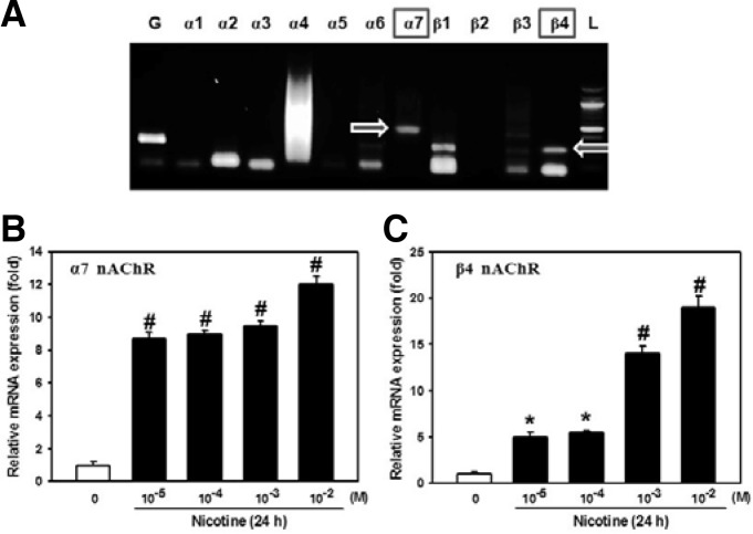
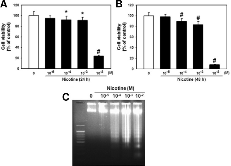
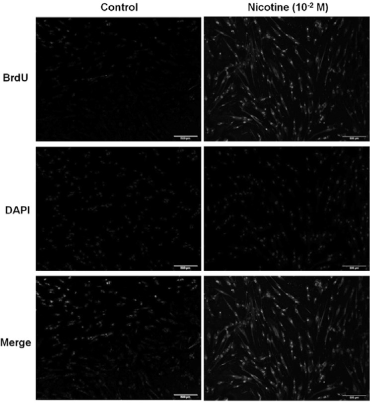
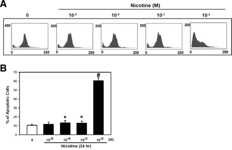
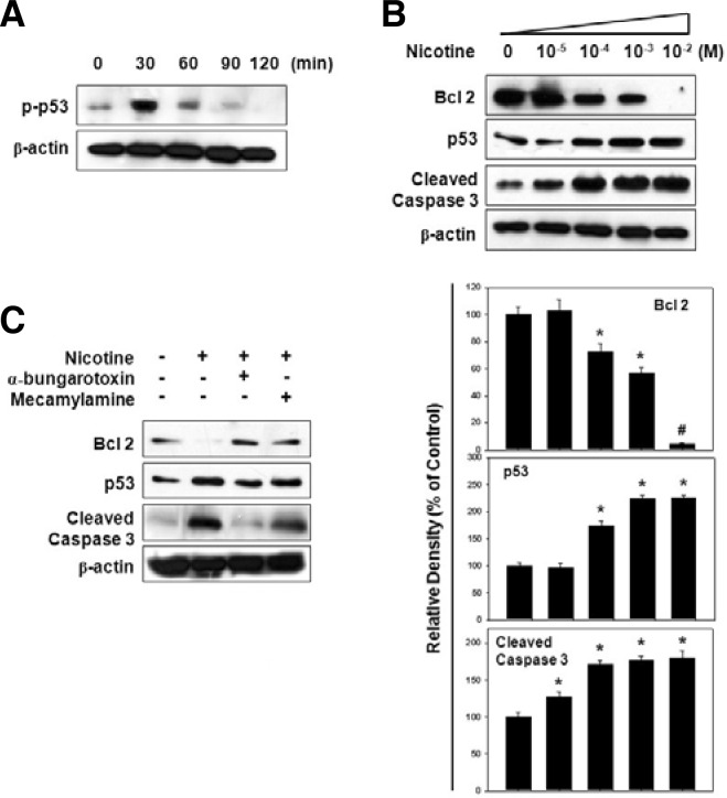
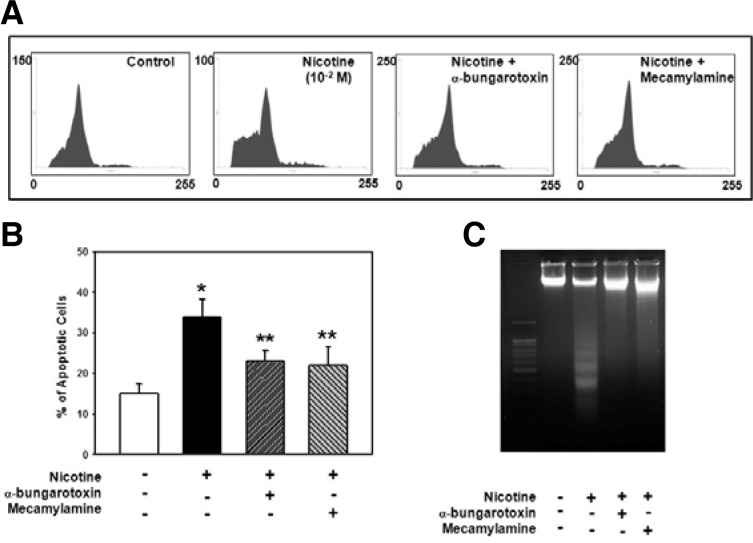
Nicotine, a major component of cigarette smoking, is the important risk factor for the development of periodontal disease. However, the mechanisms that underlie the cytotoxicity of nicotine in human periodontal ligament stem cells (PDLSCs) are largely unknown. Thus, the purpose of this study was to determine the cytotoxic effect of nicotine by means of nicotinic acetylcholine receptor (nAChR) activation in PDLSCs. We first detected α7 and β4 nAChRs in PDLSCs. The gene expressions of α7 and β4 nAChR were increased by nicotine administration. Nicotine significantly decreased cell viability at a concentration higher than 10(-5) M. DNA fragmentation was also detected at high doses of nicotine treatment. Moreover, the detection of sub G1 phase and TUNEL assay demonstrated that nicotine significantly induced apoptotic cell death at 10(-2) M concentration. Western blot analysis confirmed that p53 proteins were phosphorylated by nicotine. Under various doses of nicotine, a decrease in the anti-apoptotic protein Bcl-2, but an increase in p53 and cleaved caspase-3 protein levels, was detected in a dose-dependent manner. However, the apoptotic effect of nicotine was inhibited by the pretreatment of α-bungarotoxin, a selective α7 nAChR antagonist or mecamylamine, a non-selective nAChR antagonist. Finally, increases in the subG1 phase and DNA fragmentation by nicotine was attenuated by each nAChR antagonist. Collectively, the presence of α7 and β4 nAChRs in PDLSCs supports a key role of nAChRs in the modulation of nicotine-induced apoptosis.
Figures
References
-
- Arredondo J., Hall L.L., Ndoye A., Nguyen V.T., Chernyavsky A.I., Bercovich D., Orr-Urtreger A., Beaudet A.L., Grando S.A. Central role of fibroblast alpha3 nicotinic acetylcholine receptor in mediating cutaneous effects of nicotine. Lab. Invest. 2003;83:207–225. - PubMed
-
- Arredondo J., Chernyavsky A.I., Jolkovsky D.L., Pinkerton K.E., Grando S.A. Receptor-mediated tobacco toxicity: acceleration of sequential expression of alpha5 and alpha7 nicotinic receptor subunits in oral keratinocytes exposed to cigarette smoke. FASEB. J. 2008;22:1356–1368. - PubMed
-
- Bergstrϕm J. Cigarette smoking as risk factor in chronic periodontal disease. Community Dent. Oral Epidemiol. 1989;17:245–247. - PubMed
-
- Bergstrϕm J. Tobacco smoking and chronic destructive periodontal disease. Odontology. 2004;92:1–8. - PubMed
-
- Bosshardt D.D., Sculean A. Does periodontal tissue regeneration really work? Periodontol. 2009;2000;51:208–219. - PubMed
Publication types
MeSH terms
Substances
LinkOut - more resources
Full Text Sources
Medical
Research Materials
Miscellaneous
