Comparative Study
. 2013 Jun;35(3):583-96.
doi: 10.1007/s11357-012-9388-8. Epub 2012 Mar 3.

Accumulated hippocampal formaldehyde induces age-dependent memory decline

Affiliations
Comparative Study

Accumulated hippocampal formaldehyde induces age-dependent memory decline

Zhiqian Tong et al. Age (Dordr). 2013 Jun.

Abstract

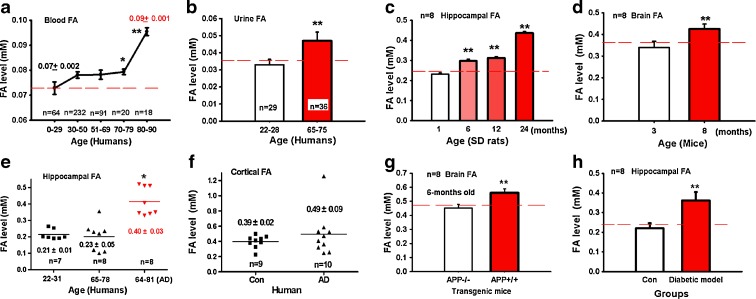
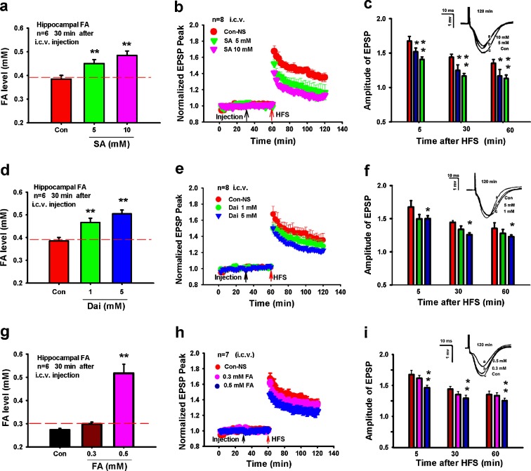
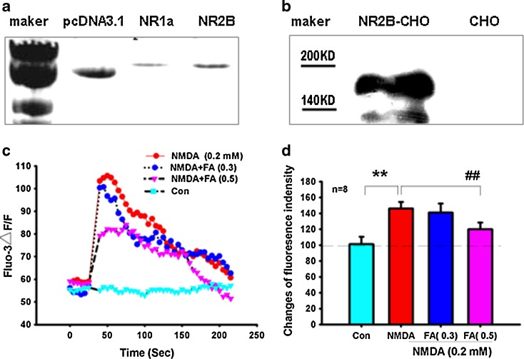
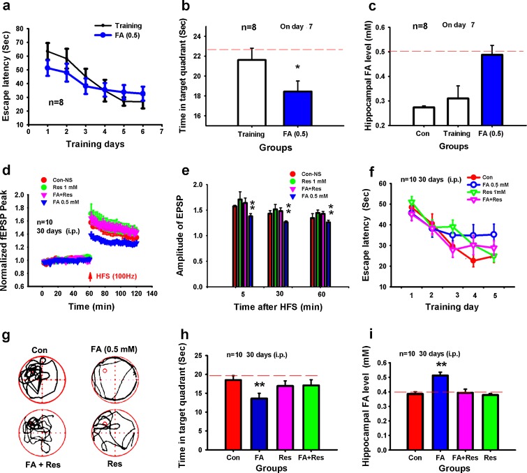
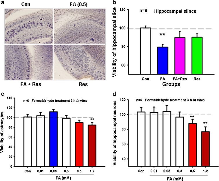
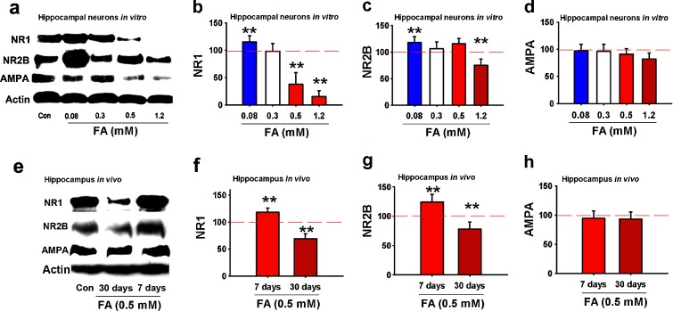
Aging is an important factor in memory decline in aged animals and humans and in Alzheimer's disease and is associated with the impairment of hippocampal long-term potentiation (LTP) and down-regulation of NR1/NR2B expression. Gaseous formaldehyde exposure is known to induce animal memory loss and human cognitive decline; however, it is unclear whether the concentrations of endogenous formaldehyde are elevated in the hippocampus and how excess formaldehyde affects LTP and memory formation during the aging process. In the present study, we report that hippocampal formaldehyde accumulated in memory-deteriorating diseases such as age-related dementia. Spatial memory performance was gradually impaired in normal Sprague-Dawley rats by persistent intraperitoneal injection with formaldehyde. Furthermore, excess formaldehyde treatment suppressed the hippocampal LTP formation by blocking N-methyl-D-aspartate (NMDA) receptor. Chronic excess formaldehyde treatment over a period of 30 days markedly decreased the viability of the hippocampus and down-regulated the expression of the NR1 and NR2B subunits of the NMDA receptor. Our results indicate that excess endogenous formaldehyde is a critical factor in memory loss in age-related memory-deteriorating diseases.


Figures

Fig. 1
Fig. 1
Endogenous formaldehyde is accumulated during aging and in some memory-deteriorating diseases. Formaldehyde levels in human blood (a) and urine (b) as aging. Formaldehyde levels in the hippocampus of SD rats (c), mouse brain (d); those in the hippocampus (e) and cortex (f) of patients with Alzheimer’s disease; and those in the brains of APP transgenic (hypomethylation of the APP gene) mice (g), and hippocampi of streptozotocin-induced diabetic SD rats (h). *P < 0.5; **P < 0.01
Fig. 2
Fig. 2
Excess endogenous formaldehyde suppresses the hippocampal LTP formation in vivo. Formaldehyde levels in hippocampi from rats treated with succinic acid (SA, an ADH3 inhibitor, i.c.v.) after 30 min (a). Effects of succinic acid on LTP (b). Statistical analyses of EPSP amplitude (c). Formaldehyde levels in hippocampi from rats treated with daidzin (Dai, an ALDH2 inhibitor, i.c.v.) after 30 min (d). Effects of daidzin on LTP (e). Statistical analyses of EPSP amplitude (f). Formaldehyde levels in hippocampi after these rats were intracerebroventricularly treated with excess formaldehyde for 30 min (g). Excess formaldehyde induces LTP suppression (h). Statistical analysis of EPSP amplitude (i). *P < 0.5; **P < 0.01
Fig. 3
Fig. 3
Excess formaldehyde non-specifically blocks the NMDA receptor in vitro. DNA bands of pcDNA3.1, NR1a, and NR2B (a). NR2B expression detected by western blotting (b). Effect of NMDA (0.2 mM) or NMDA and formaldehyde (0, 0.3, and 0.5 mM) on the [Ca2+]i fluorescence intensity of NR1/NR2B-transfected CHO cells (c). Statistical analysis of changes in fluorescence intensity (d). **P < 0.01 versus control group, ##P < 0.01 versus NMDA (0.2 mM) treatment group
Fig. 4
Fig. 4
Acute or chronic excess formaldehyde impairs spatial memory in rats. Escape latency in controls and rats injected with formaldehyde for 7 days (a). Time (seconds) spent in the target quadrant during probe trials (b). Formaldehyde levels in hippocampi from rats intraperitoneally treated with excess formaldehyde on day 7 (c). An exogenous supply of the formaldehyde scavenger resveratrol rescued LTP suppression by formaldehyde injection (i.p.) over 30 consecutive days (d). Statistical analysis of EPSP amplitude (e). Escape time (seconds) in these four groups (f). The swimming track and the time (seconds) spent in the target quadrant during probe trials (g, h). Formaldehyde levels in the hippocampus of rats intraperitoneally treated with different reagents on day 30 (i). *P < 0.5; **P < 0.01
Fig. 5
Fig. 5
Chronic excess formaldehyde decreases the viability of the hippocampus. Hippocampal slices from SD rats treated with different reagents (control, formaldehyde, formaldehyde with resveratrol, and resveratrol) for 30 consecutive days were stained with MTT solution (a). Statistical analysis of the optical intensity of MTT staining (b). Viability of cultured hippocampal neurons treated with different concentrations of formaldehyde (0, 0.01, 0.08, 0.3, 0.5, and 1.2 mM) in vitro (c). Viability of cultured astrocytes treated with different concentrations of formaldehyde (0, 0.01, 0.08, 0.3, 0.5, and 1.2 mM) in vitro (d). *P < 0.5; **P < 0.01
Fig. 6
Fig. 6
Formaldehyde regulates the expression of NMDA receptor subunits in vitro and in vivo. The effects of different concentrations of formaldehyde on the expression of NR1, NR2B, and AMPA receptors in primary cultured hippocampal neurons after formaldehyde treatment at different concentrations (0, 0.3, 0.5, and 1.2 mM) 3 h in vitro (a). Statistical analysis of the expression of NR1 (b), NR2B (c), and AMPA receptors (d). Expression of NR1, NR2B, and AMPA receptors in the hippocampi of rats injected with formaldehyde (0.5 mM) for 7 and 30 days (e). Statistical analysis of the expression of NR1 (f), NR2B (g), and AMPA receptors (h). N = 6; *P < 0.5; **P < 0.01

References

    1. Abu-Abeeleh M, Bani Ismail ZA, Alzaben KR, Abu-Halaweh SA, Aloweidi AS, Al-Ammouri IA, Al-Essa MK, Jabaiti SK, Abu-Abeeleh J, Alsmady MM. A preliminary study of the use of human adipose tissue-derived stem cells for the treatment of streptozotocin-induced diabetes mellitus in a rat model. Comp Clin Pathol. 2010;19(1):1–4. doi: 10.1007/s00580-009-0912-x. - DOI
    1. Almaguer W, Estupiņán B, Uwe Frey J, Bergado JA. Aging impairs amygdala–hippocampus interactions involved in hippocampal LTP. Neurobiology of aging. 2002;23(2):319–324. doi: 10.1016/S0197-4580(01)00278-0. - DOI - PubMed
    1. Amada N, Aihara K, Ravid R, Horie M. Reduction of NR1 and phosphorylated Ca2+/calmodulin-dependent protein kinase II levels in Alzheimer's disease. Neuroreport. 2005;16(16):1809. doi: 10.1097/01.wnr.0000185015.44563.5d. - DOI - PubMed
    1. Asaka Y, Jugloff DGM, Zhang L, Eubanks JH, Fitzsimonds RM. Hippocampal synaptic plasticity is impaired in the Mecp2-null mouse model of Rett syndrome. Neurobiol Dis. 2006;21(1):217–227. doi: 10.1016/j.nbd.2005.07.005. - DOI - PubMed
    1. Blevins T, Mirshahi T, Chandler LJ, Woodward JJ. Effects of acute and chronic ethanol exposure on heteromeric N-methyl-d-aspartate receptors expressed in HEK 293 cells. J Neurochem. 1997;69(6):2345–2354. doi: 10.1046/j.1471-4159.1997.69062345.x. - DOI - PubMed

Publication types

MeSH terms

Substances

LinkOut - more resources