. 2012 Jul;69(13):2217-31.
doi: 10.1007/s00018-012-0922-8. Epub 2012 Mar 1.

Airway basal cell vascular endothelial growth factor-mediated cross-talk regulates endothelial cell-dependent growth support of human airway basal cells

Affiliations

Airway basal cell vascular endothelial growth factor-mediated cross-talk regulates endothelial cell-dependent growth support of human airway basal cells

Giacomo Curradi et al. Cell Mol Life Sci. 2012 Jul.

Abstract

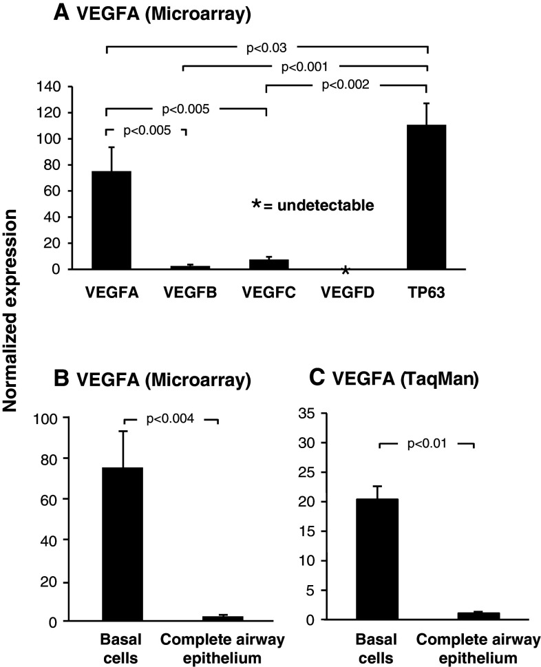
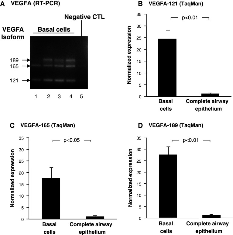
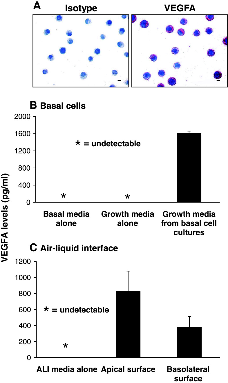
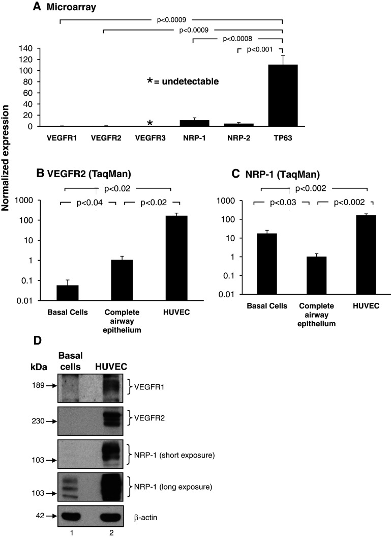
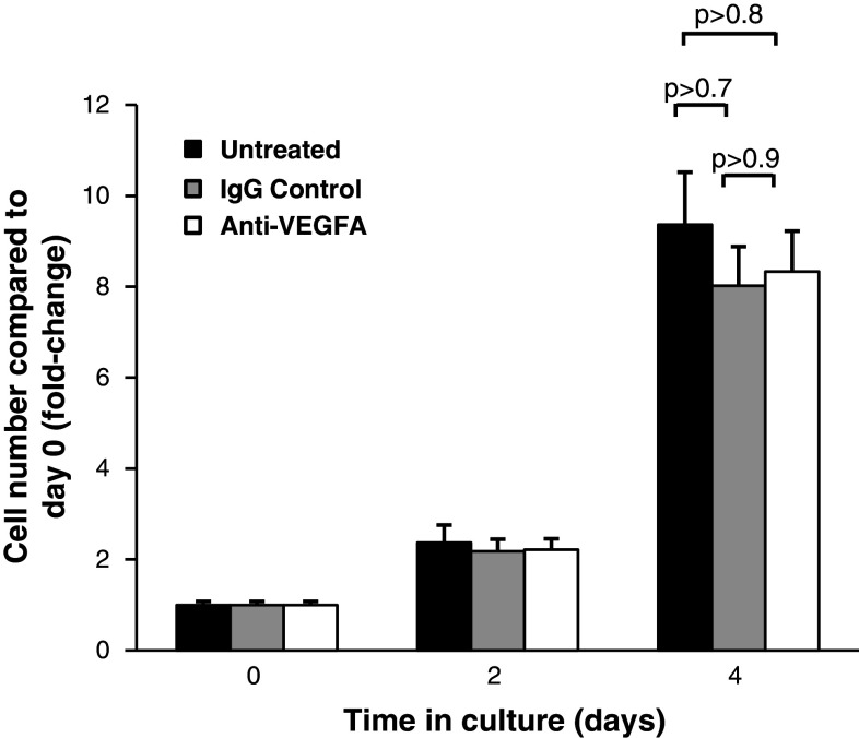
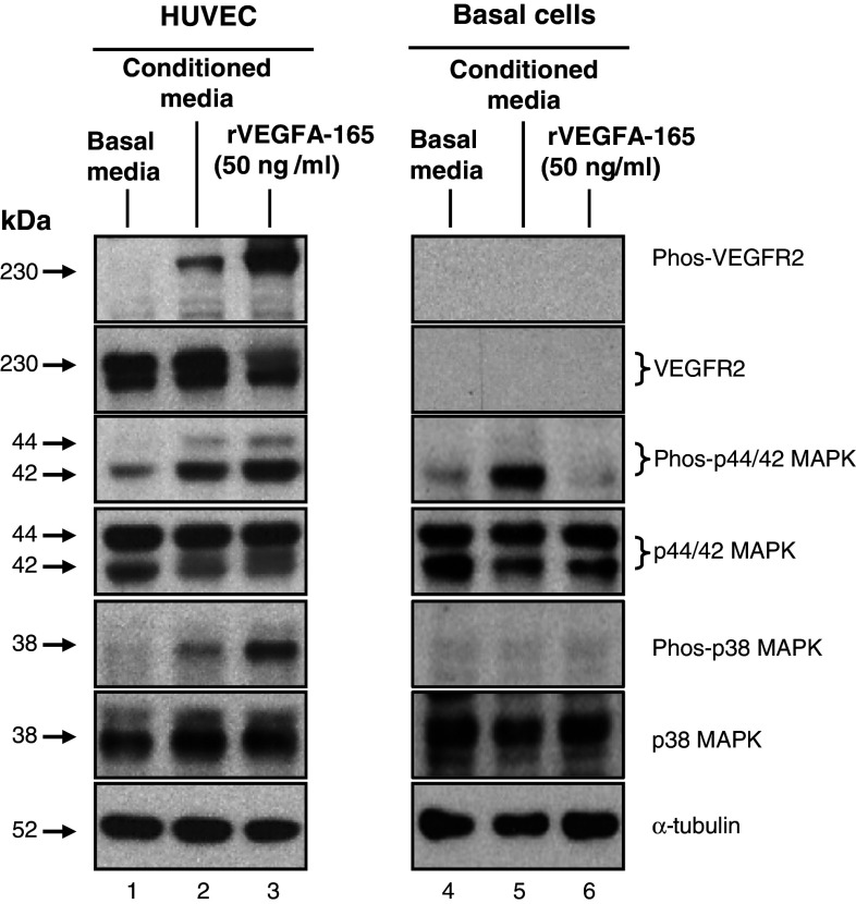
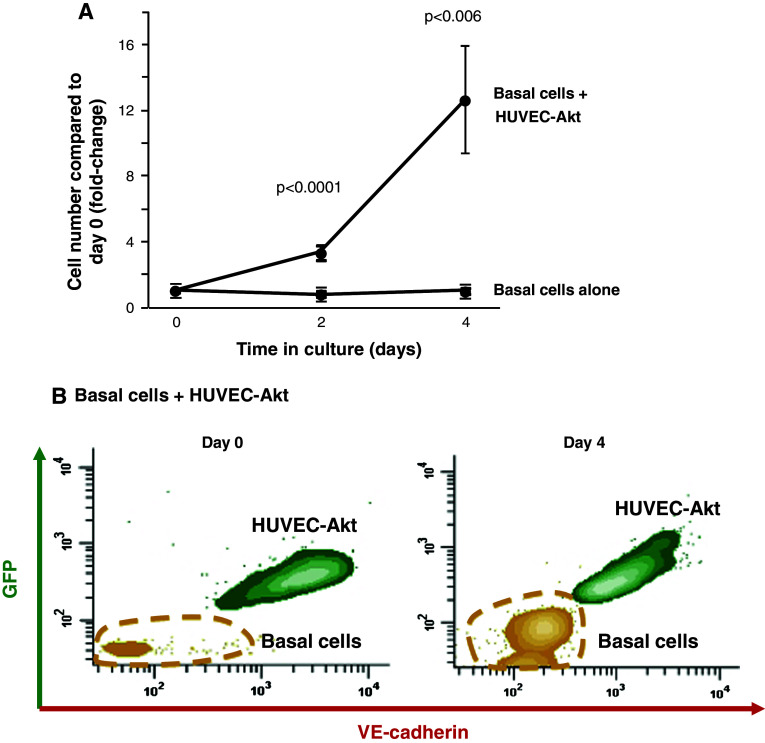
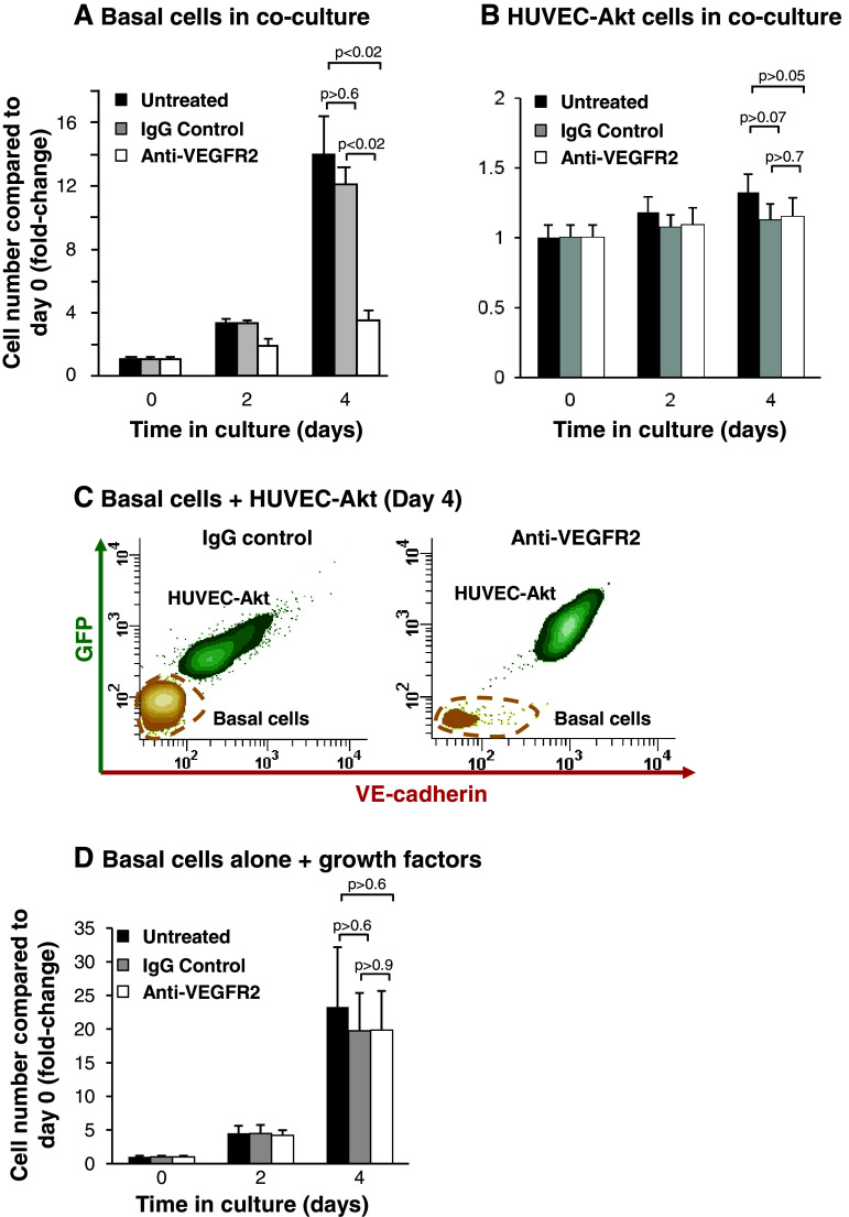
The human airway epithelium is a pseudostratified heterogenous layer comprised of ciliated, secretory, intermediate, and basal cells. As the stem/progenitor population of the airway epithelium, airway basal cells differentiate into ciliated and secretory cells to replenish the airway epithelium during physiological turnover and repair. Transcriptome analysis of airway basal cells revealed high expression of vascular endothelial growth factor A (VEGFA), a gene not typically associated with the function of this cell type. Using cultures of primary human airway basal cells, we demonstrate that basal cells express all of the three major isoforms of VEGFA (121, 165 and 189) but lack functional expression of the classical VEGFA receptors VEGFR1 and VEGFR2. The VEGFA is actively secreted by basal cells and while it appears to have no direct autocrine function on basal cell growth and proliferation, it functions in a paracrine manner to activate MAPK signaling cascades in endothelium via VEGFR2-dependent signaling pathways. Using a cytokine- and serum-free co-culture system of primary human airway basal cells and human endothelial cells revealed that basal cell-secreted VEGFA activated endothelium to express mediators that, in turn, stimulate and support basal cell proliferation and growth. These data demonstrate novel VEGFA-mediated cross-talk between airway basal cells and endothelium, the purpose of which is to modulate endothelial activation and in turn stimulate and sustain basal cell growth.


Figures

Fig. 1
Fig. 1
Vascular endothelial growth factor A is highly expressed in cultured human airway basal cells. a Microarray analysis of VEGF ligand expression VEGFA (probeset: 212171_x_at), VEGFB (probeset: 203683_s_at), VEGFC (probeset: 209946_at) and VEGFD (probset: 206742_at) in basal cells (n = 4). For comparison, expression levels of the basal-cell-specific gene TP63 (probeset: 209863_s_at) is included. b Microarray analysis of VEGFA expression (probeset: 212171_x_at) in basal cells (n = 4) compared to complete airway epithelium (n = 22). c TaqMan analysis of VEGFA expression (all isoforms) in basal cells (n = 3) compared to complete airway epithelium (n = 3)
Fig. 2
Fig. 2
Expression of VEGFA isoforms 121, 165, and 189 in cultured human airway basal cells. a PCR amplification of VEGFA isoforms in basal cells (n = 4). Lanes 1–4: examples of four independent basal cell cultures. VEGFA-121, VEGFA-165, and VEGFA-189 are expressed. Lane 5: negative control (no DNA added). bd TaqMan analysis of VEGFA isoform expression in basal cells (n = 3) compared to complete airway epithelium (n = 3) using isoform specific probes. b VEGFA-121, c VEGFA-165, and d VEGFA-189
Fig. 3
Fig. 3
Expression and secretion of VEGFA by cultured human airway basal cells. a Immunohistochemical staining of VEGFA in human airway basal cells (bar = 10 μm). b VEGFA secretion by human airway basal cells. VEGFA levels assessed by ELISA in basal media, growth media, and growth media from basal cell cultures (n = 9). c VEGFA secretion by human airway basal cells during air–liquid interface culture (ALI). VEGFA levels assessed by ELISA in ALI media, and ALI media exclusively exposed to the apical surface (upper chamber) or basolateral surface (lower chamber) of basal cells during ALI culture (n = 3)
Fig. 4
Fig. 4
Expression of VEGF receptors and co-receptors in cultured human airway basal cells. a Microarray analysis of VEGFR1 (probeset: 226497_s_at), VEGFR2 (probeset: 203934_at), VEGFR3 (probeset: 210316_at), NRP-1 (probeset: 212298_at) and NRP-2 (probeset: 229225_at) expression in basal cells (n = 4). For comparison, expression levels of the basal-cell-specific gene TP63 (probeset: 209863_s_at) are included. bc TaqMan analysis of VEGFR2 and NRP-1 expression in basal cells (n = 4) and human umbilical vein endothelial cells (HUVEC) (n = 4) compared to complete airway epithelium (n = 4) using specific probes. b VEGFR2, c NRP-1. d Western-blot analysis of VEGFR1, VEGFR2, and NRP-1 in basal cells compared to HUVEC. Lane 1: basal cells; lane 2: HUVEC. For both cell types, shown is expression of VEGFR1, VEGFR2, NRP-1 (short and long exposure) and β-actin as a loading control
Fig. 5
Fig. 5
Inhibition of VEGFA signaling has no effect on proliferation of airway basal cells. Human airway basal cells were cultured in growth media and incubated with control IgG or blocking monoclonal antibody against VEGFA. Data shown is the average of three independent experiments. Untreated (black), IgG control (gray), and anti-VEGFR2 (white)
Fig. 6
Fig. 6
Secreted VEGFA from airway basal cells activates endothelium via VEGFR2-mediated signaling. Human umbilical vein endothelial cells (HUVEC) and human airway basal cells were serum-starved for 6 h and then stimulated with basal media (without serum or cytokines), basal media conditioned with basal cells, or, as a positive control, basal media containing recombinant VEGFA-165 (50 ng/ml). Following stimulation, cell lysates were prepared and the activation of endothelium and basal cells was evaluated by Western-blot analysis and staining for phosphorylated VEGFR2 (Phos-VEGFR2), phosphorylated p44/42 MAPK (Phos-p44/44 MAPK) and phosphorylated p38 MAPK (Phos-p38 MAPK). The levels of total VEGFR2, p44/42 MAPK, and p38 MAPK were also evaluated. α-tubulin was used as a loading control. Lane 1: lysates of HUVEC exposed to basal cell media alone; lane 2: HUVEC exposed to conditioned media; lane 3: recombinant VEGFA-165; lanes 4–6: identical to lanes 1–3, but with lysates of basal cells
Fig. 7
Fig. 7
Endothelial cells support the growth of airway basal cells in the absence of growth factors. a Human airway basal cells were cultured alone or in co-culture with Akt-activated human umbilical vein endothelial cells (HUVEC-Akt) in cytokine- and serum-free conditions. At the desired time points, cells were harvested and the GFP-labeled HUVEC-Akt cells was determined as the GFP+VE-cadherin+ population by flow cytometric analysis, and the GFPVE-cadherin population quantified as expanded basal cells. Data shown is the average of four independent experiments. b Representative flow cytometric analysis of human airway basal cell and HUVEC-Akt populations at day 0 and day 4 of co-culture. HUVEC-Akt cells were determined as the GFP+VE-cadherin+ population, and the GFPVE-cadherin population quantified as expanded basal cells
Fig. 8
Fig. 8
Inhibition of VEGFR2 signaling suppresses endothelial cell-dependent proliferation of airway basal cells. ab Human airway basal cells were co-cultured with human umbilical vein endothelial cells activated with Akt (HUVEC-Akt) in cytokine- and serum-free conditions and incubated with control IgG or blocking monoclonal antibodies against VEGFR2. Data shown is the average of three independent experiments. a Basal cells, b HUVEC-Akt cells, c representative flow cytometric analysis of human airway basal cell and HUVEC-Akt cell populations at day 4 of co-culture following incubation with control IgG or anti-VEGFR2. HUVEC-Akt cells were determined as the GFP+VE-cadherin+ population, and the GFPVE-cadherin population quantified as basal cells, and d human airway basal cells were cultured in growth media and incubated with control IgG or blocking monoclonal antibodies against VEGFR2. Data shown is the average of three independent experiments. For all panels, shown are untreated (black), IgG control (gray), and anti-VEGFR2 (white)

References

    1. Breeze RG, Wheeldon EB. The cells of the pulmonary airways. Am Rev Respir Dis. 1977;116:705–777. - PubMed
    1. Mercer RR, Russell ML, Roggli VL, Crapo JD. Cell number and distribution in human and rat airways. Am J Respir Cell Mol Biol. 1994;10:613–624. - PubMed
    1. Knight DA, Holgate ST. The airway epithelium: structural and functional properties in health and disease. Respirology. 2003;8:432–446. doi: 10.1046/j.1440-1843.2003.00493.x. - DOI - PubMed
    1. Thompson AB, Robbins RA, Romberger DJ, Sisson JH, Spurzem JR, Teschler H, et al. Immunological functions of the pulmonary epithelium. Eur Respir J. 1995;8:127–149. doi: 10.1183/09031936.95.08010127. - DOI - PubMed
    1. Crystal RG, Randell SH, Engelhardt JF, Voynow J, Sunday ME. Airway epithelial cells: current concepts and challenges. Proc Am Thorac Soc. 2008;5:772–777. doi: 10.1513/pats.200805-041HR. - DOI - PMC - PubMed

Publication types

MeSH terms

LinkOut - more resources