. 2012 Feb 29;483(7387):59-63.
doi: 10.1038/nature10883.

Recognition of SUMO-modified PCNA requires tandem receptor motifs in Srs2

Affiliations

Recognition of SUMO-modified PCNA requires tandem receptor motifs in Srs2

Anthony A Armstrong et al. Nature. .

Abstract

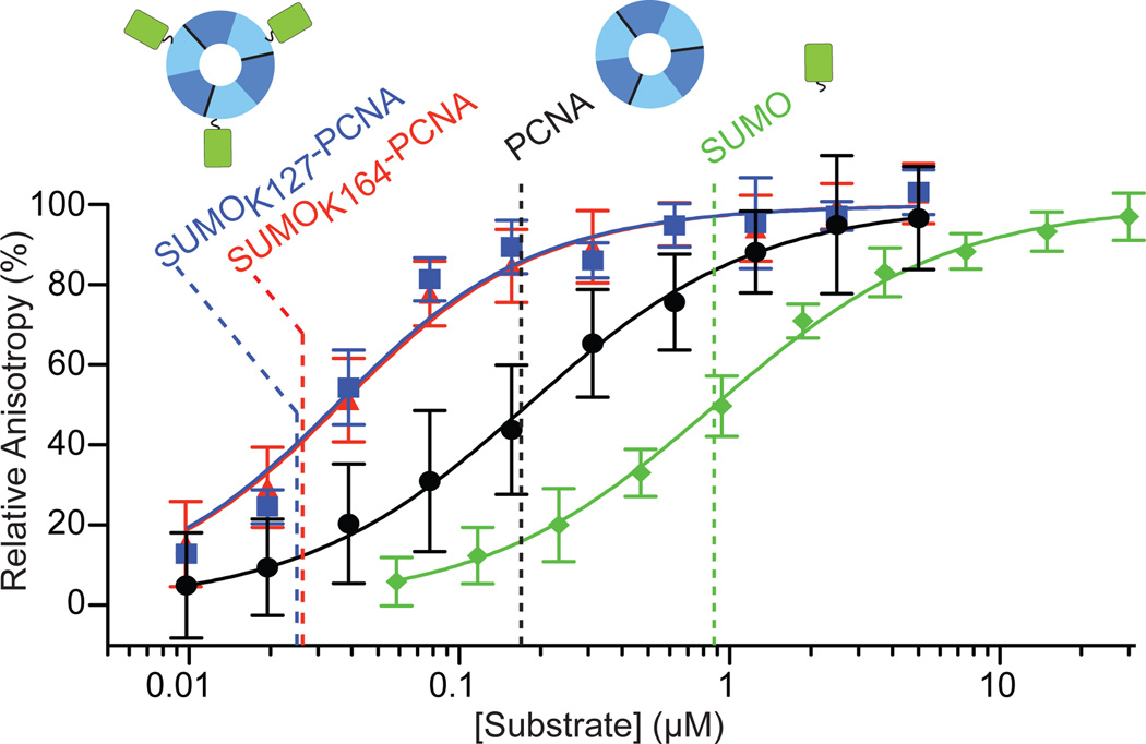
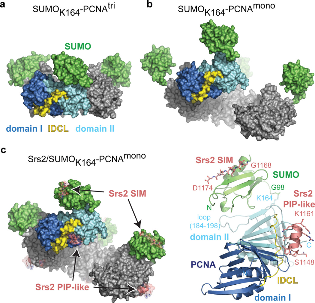
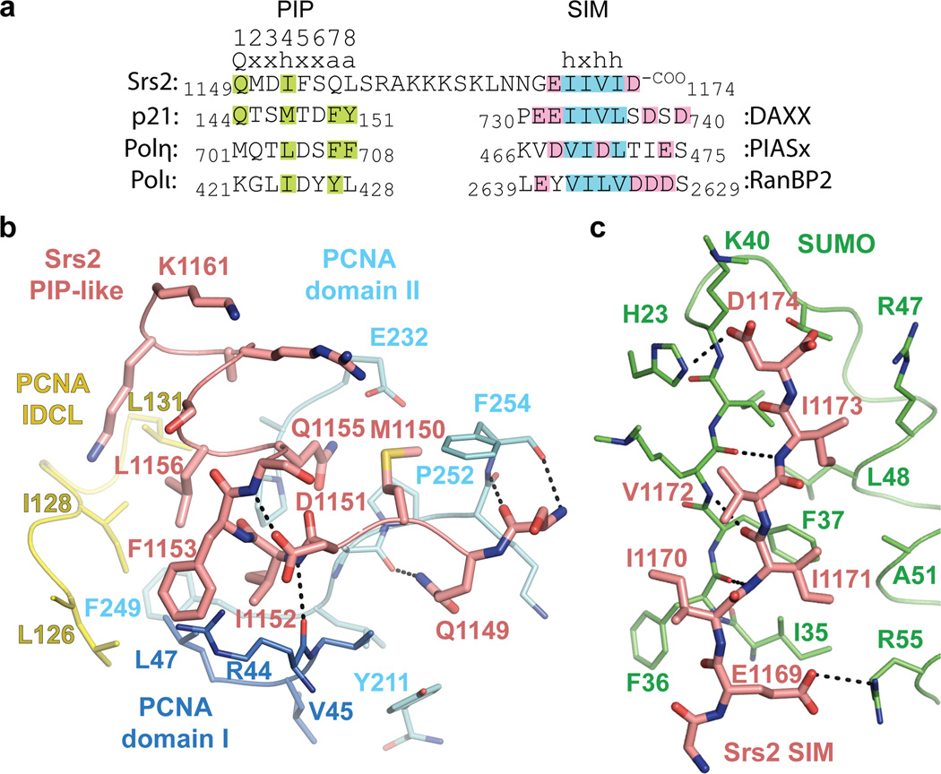
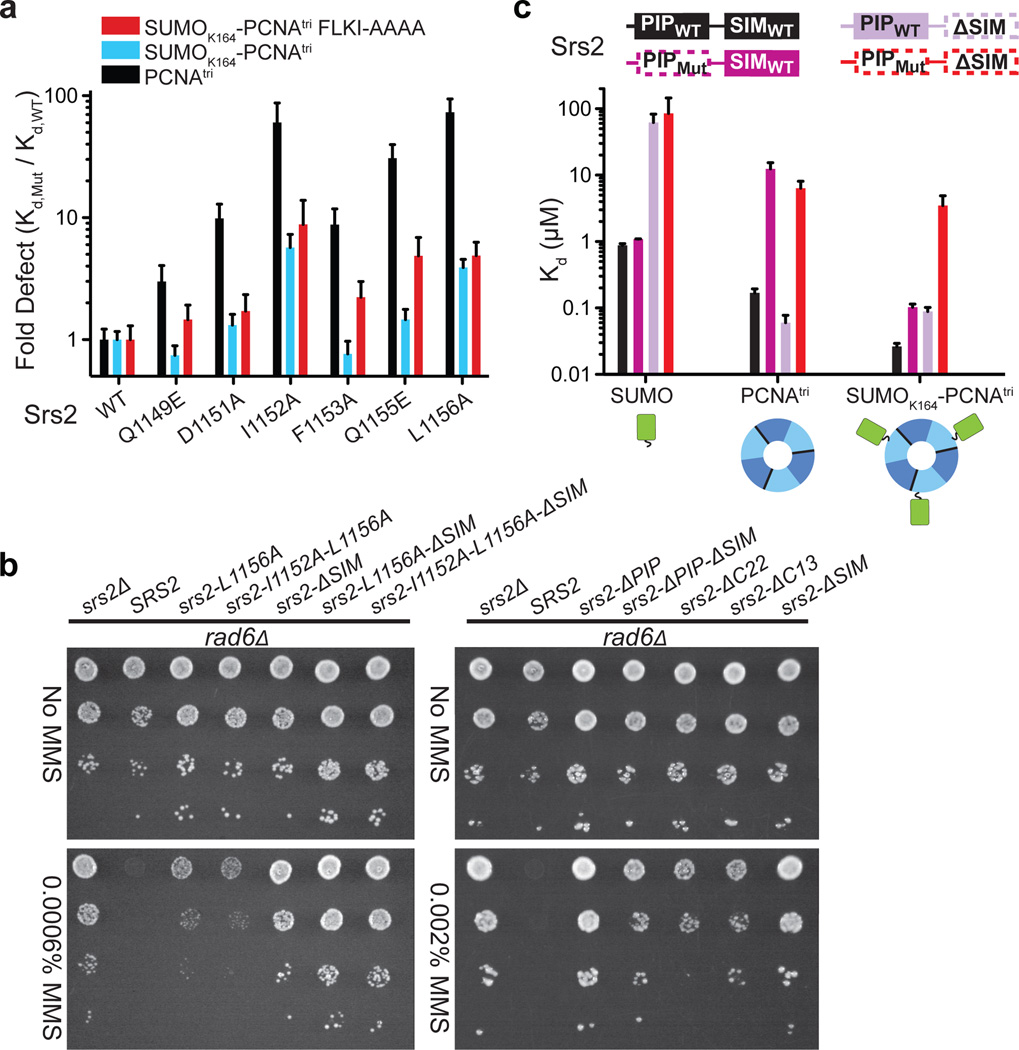
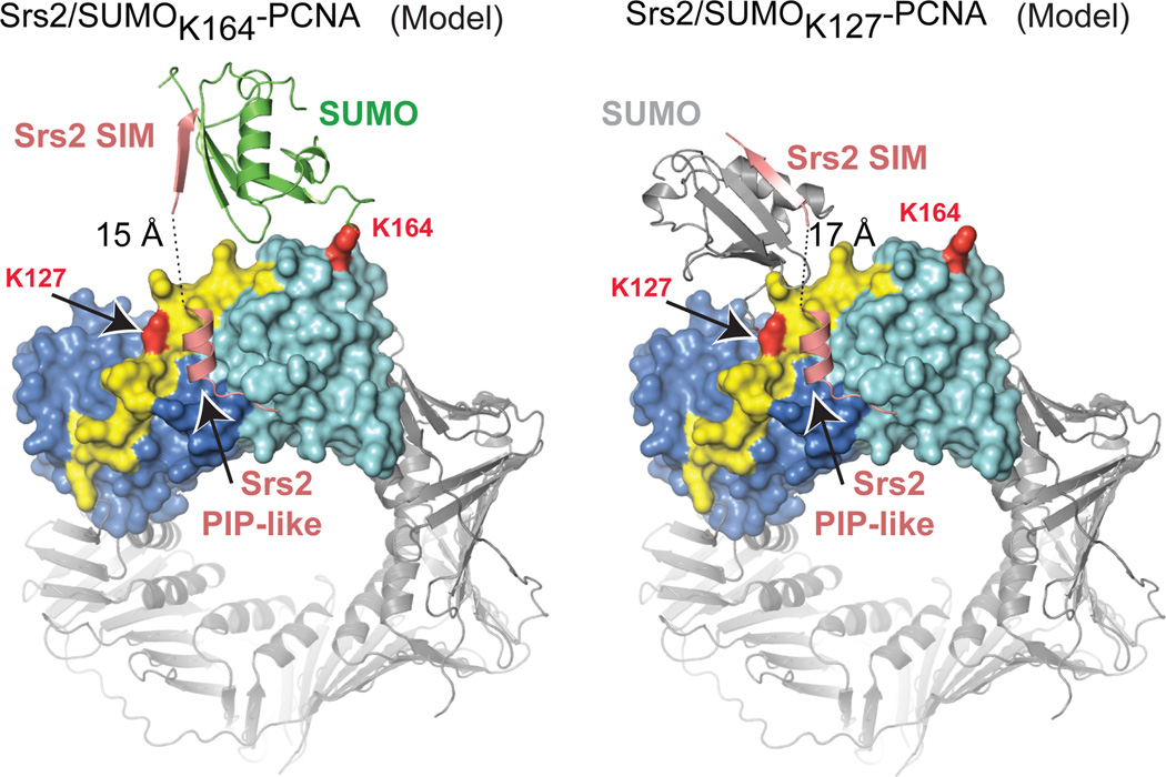
Ubiquitin (Ub) and ubiquitin-like (Ubl) modifiers such as SUMO (also known as Smt3 in Saccharomyces cerevisiae) mediate signal transduction through post-translational modification of substrate proteins in pathways that control differentiation, apoptosis and the cell cycle, and responses to stress such as the DNA damage response. In yeast, the proliferating cell nuclear antigen PCNA (also known as Pol30) is modified by ubiquitin in response to DNA damage and by SUMO during S phase. Whereas Ub-PCNA can signal for recruitment of translesion DNA polymerases, SUMO-PCNA signals for recruitment of the anti-recombinogenic DNA helicase Srs2. It remains unclear how receptors such as Srs2 specifically recognize substrates after conjugation to Ub and Ubls. Here we show, through structural, biochemical and functional studies, that the Srs2 carboxy-terminal domain harbours tandem receptor motifs that interact independently with PCNA and SUMO and that both motifs are required to recognize SUMO-PCNA specifically. The mechanism presented is pertinent to understanding how other receptors specifically recognize Ub- and Ubl-modified substrates to facilitate signal transduction.


Figures

Figure 1
Figure 1. Srs2 C-terminal domain interacts with PCNA, SUMO, and SUMO-PCNA
Fluorescence anisotropy curves and calculated Kd values (dashed lines) for GST-SUMO (green), PCNA (black), SUMOK127-PCNA (blue) and SUMOK164-PCNA (red) titrations against 20 nM BODIPY-FL Srs21107–1174. Cartoons of SUMO (green; right), PCNA (trimer, domain I (dark blue), domain II (light blue); middle) and SUMO-PCNA (left) above dashed lines. Assays conducted in triplicate. Error bars are ±1 standard deviation; n = 12 for SUMO, n = 12 for PCNA, n = 9 for SUMOK164-PCNA and n= 3 for SUMOK127-PCNA. See Methods.
Figure 2
Figure 2. Structures of SUMO-PCNA alone and in complex with Srs2
a, Surface representation for SUMOK164-PCNAtri. PCNA domain I (dark blue), domain II (light blue), IDCL (yellow) and SUMO (green) are labeled. b, SUMOK164-PCNAmono as in a. Four protomers oriented to visualize the ‘open ring’. c, Srs21107–1174/SUMOK164-PCNAmono as in a and b (left). Srs2 SIM and PIP-like motifs labeled (pink; stick and transparent surface). Cartoon representation for one protomer of Srs21107–1174/SUMOK164-PCNAmono (right) colored as in c. PCNA N- and C- termini, loop 184–198, and K164 and SUMO N-terminus and G98 are labeled. Srs2 SIM and PIP-like motifs labeled with single amino acid code and number to indicate the termini of each motif.
Figure 3
Figure 3. Srs2 PIP-like motif and SIM interactions with PCNA and SUMO
a, Sequence alignment for Srs2 CTD (amino acids 1149–1174) above p21, Polη and Polι PIP-box motifs and DAXX, PIASx and RanBP2 SIMs. PIP-box consensus motif numbered above Srs2 (Q=glutamine, x=any amino acid, h=hydrophobic, a=aromatic). Amino acids similar to consensus are highlighted (green). Hydrophobic and acidic residues highlighted blue and pink for SIMs. b, Srs2 PIP-like motif (pink), PCNA colored as in Fig 2. PCNA, Srs2 and select amino acids labeled by numbered single letter code. Hydrogen bonds as dashed lines. c, Srs2 SIM (pink) and SUMO (green) as in b.
Figure 4
Figure 4. Srs2 PIP-like motif and SIM required for recognition of SUMO-PCNA
a, Bar graphs showing fold-defect in Kd (y-axis) for WT and Srs21107–1174 mutants (x-axis) for PCNAtri K127G (black), SUMOK164-PCNAtri K127G (blue), or SUMOK164-PCNAtri FLKI125–128AAAA (red). b, Suppression of rad6Δ DNA damage sensitivity by srs2Δ or srs2 alleles. Shown are 10-fold serial dilutions of cells on plates containing MMS as indicated (left). c, Bar graphs showing Kd values for interaction of Srs2 containing a wild-type PIP and SIM (PIPWT-SIMWT) (black), a mutant PIP-like motif (L1156A; PIPMut) and SIMWT (dark purple; dashed line around PIPMut), a PIPWT with SIM deletion (ΔSIM) (light purple; dashed line around ΔSIM), and a PIPMut ΔSIM (red dashed line) with SUMO (left), PCNA (middle) and SUMOK164-PCNAtri (right). Cartoons colored as in Fig. 1. FP assays conducted in triplicate. Error bars are ±1 standard deviation. See Methods.
Figure 5
Figure 5. Models for Srs2/SUMO-PCNA complexes
Models for SUMO conformations to enable simultaneous interaction with the SIM and PIP-like motif when attached to PCNA K164 (left) or K127 (right). Models generated required a simple rotation of SUMO at the isopeptide linkage for SUMOK164-PCNA and a slight rotation and translation of SUMO from a symmetry related complex to mimic SUMOK127-PCNA (Supplemental Fig. 13).

References

    1. Kirkin V, Dikic I. Role of ubiquitin- and Ubl-binding proteins in cell signaling. Curr Opin Cell Biol. 2007;19:199–205. - PubMed
    1. Gareau JR, Lima CD. The SUMO pathway: emerging mechanisms that shape specificity, conjugation and recognition. Nat Rev Mol Cell Biol. 2010;11:861–871. - PMC - PubMed
    1. Dikic I, Wakatsuki S, Walters KJ. Ubiquitin-binding domains - from structures to functions. Nat Rev Mol Cell Biol. 2009;10:659–671. - PMC - PubMed
    1. Moldovan GL, Pfander B, Jentsch S. PCNA, the maestro of the replication fork. Cell. 2007;129:665–679. - PubMed
    1. Krishna TS, Kong XP, Gary S, Burgers PM, Kuriyan J. Crystal structure of the eukaryotic DNA polymerase processivity factor PCNA. Cell. 1994;79:1233–1243. - PubMed

Methods References

    1. Mossessova E, Lima CD. Ulp1-SUMO crystal structure and genetic analysis reveal conserved interactions and a regulatory element essential for cell growth in yeast. Mol Cell. 2000;5:865–876. - PubMed
    1. Rayment I. Reductive alkylation of lysine residues to alter crystallization properties of proteins. Methods Enzymol. 1997;276:171–179. - PubMed
    1. Otwinowski Z, Minor W. In: Methods Enzymol. Carter CW Jr, Sweet RM, editors. Vol. 276. Academic Press; 1997. pp. 307–326. - PubMed
    1. Collaborative Computational Project. The CCP4 suite: programs for protein crystallography. Acta Crystallogr D Biol Crystallogr. 1994;50:760–763. - PubMed
    1. Vagin A, Teplyakov A. MOLREP: an automated program from molecular replacement. J Appl Cryst. 1997;30:1022–1025.

Publication types

MeSH terms

Substances