. 2012 Jun;13(3):335-49.
doi: 10.1007/s10162-012-0316-5. Epub 2012 Mar 2.

Head bobber: an insertional mutation causes inner ear defects, hyperactive circling, and deafness

Affiliations

Head bobber: an insertional mutation causes inner ear defects, hyperactive circling, and deafness

Giuseppina Somma et al. J Assoc Res Otolaryngol. 2012 Jun.

Abstract

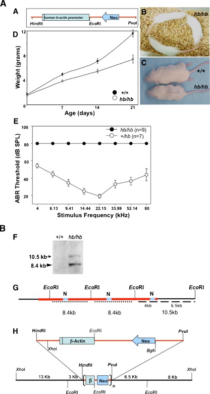
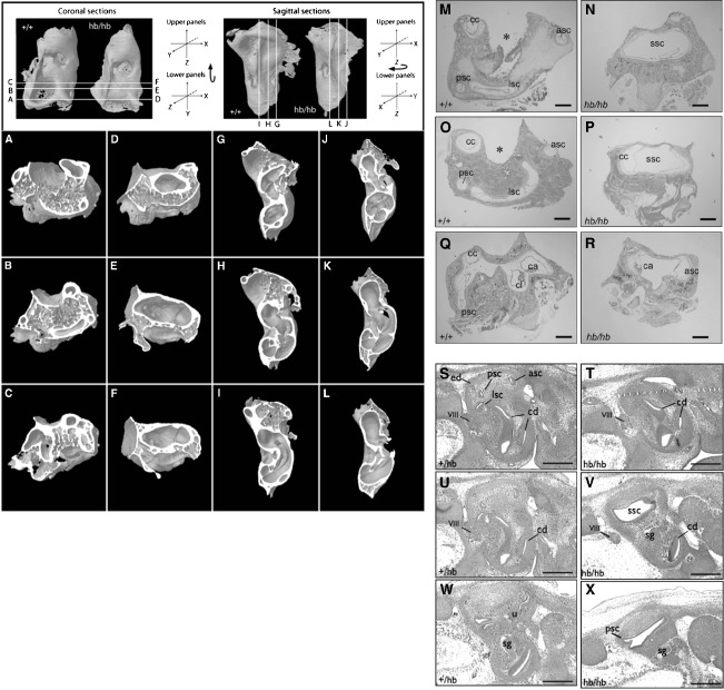
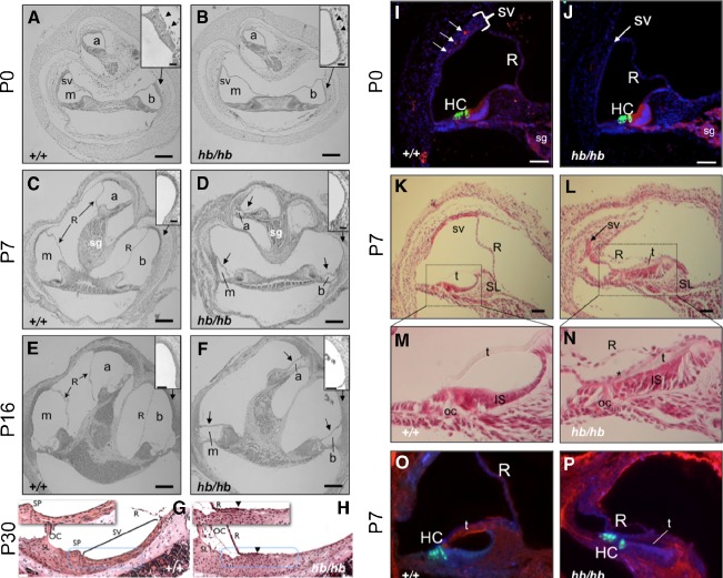
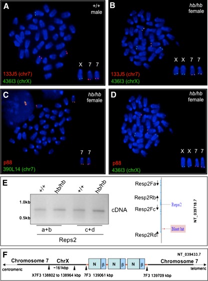
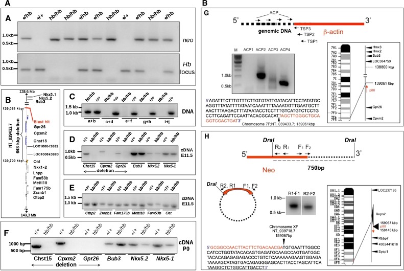
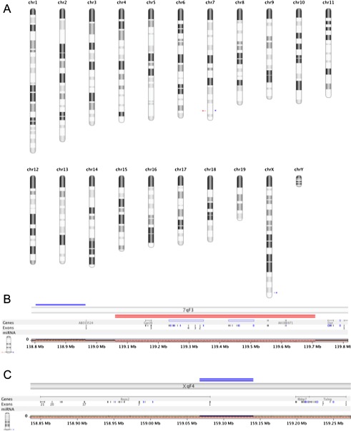
The head bobber transgenic mouse line, produced by pronuclear integration, exhibits repetitive head tilting, circling behavior, and severe hearing loss. Transmitted as an autosomal recessive trait, the homozygote has vestibular and cochlea inner ear defects. The space between the semicircular canals is enclosed within the otic capsule creating a vacuous chamber with remnants of the semicircular canals, associated cristae, and vestibular organs. A poorly developed stria vascularis and endolymphatic duct is likely the cause for Reissner's membrane to collapse post-natally onto the organ of Corti in the cochlea. Molecular analyses identified a single integration of ~3 tandemly repeated copies of the transgene, a short duplicated segment of chromosome X and a 648 kb deletion of chromosome 7(F3). The three known genes (Gpr26, Cpxm2, and Chst15) in the deleted region are conserved in mammals and expressed in the wild-type inner ear during vestibular and cochlea development but are absent in homozygous mutant ears. We propose that genes critical for inner ear patterning and differentiation are lost at the head bobber locus and are candidate genes for human deafness and vestibular disorders.


Figures

FIG. 1
FIG. 1
A, B Phenotype and molecular characterization of the genomic region interrupted by the transgene insertion in head bobber mice. A Diagram of the transgene fragment used to create head bobber line. B A composite of three images showing hb/hb mice display hyperactivity and circling behavior. C hb/hb mice are reduced size in adulthood and have a reduced growth compared with wild-type littermates (D). Black bar in human β-actin promoter represents exon 1. E Auditory brainstem response (ABR) thresholds were measured for hemizygotes (+/hb, n = 7) and homozygotes (hb/hb, n = 9) at P20. +/hb had normal hearing thresholds, but hb/hb mice had no responses at any frequencies tested. Data are presented as mean ± SEM in D and E. F Southern analysis of the hb locus with a neo gene probe (grey box, N in G) hybridized to two bands (10.5 and 8.4 kb) indicating a single insertion site (red line) of the transgene in the head bobber genome. The 11 kb band represents a single-copy transgene plus flanking genomic sequences (dashed line in G) and the 8.4 kb band represents the multicopy concatamer (dotted line in G). Appropriately, no bands were detected in the wild-type (wt) genome. G Restriction map of the transgene vector and genome allowed the creation of a map (H) of the genomic region surrounding the transgene integration site. β, human β-actin gene fragment; black line in human β-actin gene represents exon 1; n, multicopy concatamer. dB, decibels; SPL, sound pressure levels; kHz, kilohertz.
FIG. 2
FIG. 2
Head bobber defects in semicircular canal and cochlear development. Micro-computed tomography coronal (AF) and sagittal sections (I–J) of inner ears from P20 +/+ (A–C; G–I) and hb/hb (D–F; J–L), transverse sections from P7 +/+ (M–Q) and hb/hb (N–R) and E13 +/+ (S, U, W) and hb/hb (T, V, X) mice showing inner ear defects in hb/hb mice. hb/hb mice show severe malformation of the semicircular canal region (ssc): the anterior (asc), the posterior (psc), and lateral (lsc) canals, and crista are absent presenting a single vacuous chamber (D–F, J–L, and N,P). Panel R shows a reduced canal (presumed the anterior) with an associated crista. The absence of the SSCs is evident in hb/hb at E13 (compare V to U). cc, crus commune; ca, anterior canal crista; cd, cochlear duct; cl, lateral canal crista; ed, endolymphatic duct; sg, spiral ganglion; u, utricle; VIII, eighth cranial nerve. Scale bar = 100 μm (M–X).
FIG. 3
FIG. 3
Defects in the stria and altered differentiation in the cochlea of head bobber ears. Transverse sections of inner ears are shown from P0 (A–B; I–J), P7 (C–D; K–P), P16 (E–F), and P30 (G–H), +/+ (A–O) and hb/hb (B–P). hb/hb have defects in the scala media. Sections through the cochlea show a poorly developed stria vascularis (sv, compare arrowheads in A and B insets) in hb/hb at P0. The scala media is further compromised by a collapse of Reissner’s membrane (R), shown at P7 (arrows in D) and P16 (arrows in F). The stria vascularis has completely lost the multilayer structure and appearing flat with reduced capillaries (compare insets in C to D and panels G to H). Moreover, blood vessels are readily visualized in wild-type (I) but severely reduced in the stria (sv) of hb/hb ears (J) as detected by anti-cadherin immunostaining (red) at P0. The stria in hb/hb (arrow in J) is significantly thinned compared with the DAPI-stained nuclei in the wild-type (bracketed in I). In the organ of Corti (oc), hb/hb hair cells (HC) and spiral ganglion (SG) express appropriate differentiation markers; compare myosin VII (green) and cadherin (red) in J to I. At P7, the hb/hb stria (arrow in L) is a remnant compared with the wild-type (K) stria at the apical turn and Reissner’s (R), and tectorial (t) membranes are collapsed onto the poorly differentiated inner sulcus (IS) (compare K to L, M to N and O to P). There also is a reduced expression of fibronectin (blue) in the hb/hb tectorial membrane (compare t in P to O). a, apical scala; b, basal scala; m, media scala; sg, spiral ganglion. GSLI lectin (red); SL, spiral limbus, SP, spiral prominence. Scale bar = 100 μm (AF); 20 μm (insets in A, B; C, D; E, F, and in panels G–H); or 50 μm (I–P).
FIG. 4
FIG. 4
The head bobber locus on chromosome 7 contains a duplicated fragment of chromosome X. A Chromosome-7- (133J5, red) and chromosome-X (436I3, green)-specific BAC clones are seen to hybridize to their respective chromosomal loci in wild-type (wt) male spleen cells, whereas in female hb/hb cells, the chromosome 7 BAC clone (436J3, red) also co-localizes with the chromosome X (133J5, green) clone (B). Both chromosome 7 (green in C) and chromosome X (green in D) BAC clones co-localize with the p88 vector on the chromosome 7 from hb/hb spleen cells. E Splice variants of the Reps2 gene are appropriately expressed in hb/hb ears as measured by RT-PCR (see “Methods” for primers used). Blast hit, sequence comparison location. F Diagram of the head bobber locus (red line) showing nucleotide positions of the transgene integration site and junctions on chromosome 7 having three copies of the transgene (green boxes, neo; blue boxes, β-actin cassette). chr7, chromosome 7; chrX, chromosome X.
FIG. 5
FIG. 5
Head bobber genotyping, delineation of the deleted portion of chromosome 7, and identification of the transgene integration site. A PCR genotyping using Neo (top) and chromosome 7 (bottom) primer sets in +/+, hemizygous (+/hb), and head bobber (hb/hb) mice. B Haplotype diagram of the head bobber locus on chromosome 7 and gene loci interrogated by PCR analysis (C) for the extent of genomic deletion surrounding the hb locus (primers a–i are listed in Table I). Blast hit in B, initial sequence comparison location. D–F RT-PCR analyses for gene expression (primers are listed in Table II) in the head bobber region at E11.5 (D, E) and at P0 (F) from +/+ and head bobber (hb/hb) inner ears. G Using specific primers (TSP1, TSP2, TSP3, top panel) designed to the β-actin region and DNA Walking primers (top panel, ACP; designed to capture transgene flanking genomic sequences) specific bands were amplified (bottom panel) that were mapped to sequences on chromosome 7 (right panel). H Inverse PCR of DraI digested hb/hb genomic DNA (top panel) was circularized (middle panel), and nested primers (reverse R1, R2, and forward F1, F2) designed in the Neo gene were used to perform primary (R1–F1) and secondary (R2–F2) inverse PCR resulting in a approx. 750 bp band (bottom panel) that was mapped to chromosome X. Sequencing of cloned fragments allowed orientation of the transgene in the genome (arrows in chromosome maps).
FIG. 6
FIG. 6
Comparative genomic analysis (aCGH) of hb/hb and +/+ genomes. A All chromosome view of significant genomic variations in the hb/hb genome showing loss (red arrowhead) and gain (blue arrowhead) in chromosome 7 and a gain (blue arrowhead) on the X chromosome. B View of chromosome 7qF3 region showing copy number variations. Single blue line depicts gain (ch7/qF3 138,802,774–138,964,543) and double red lines depict homozygous loss of genomic region encompassing chr7qF2: 139,060,510–139,709,028. C View of chromosome XqF4 showing a gain of chX/qF4 159,067,363–159,140,649.

References

    1. Ahmed ZM, Riazuddin S, Bernstein SL, Ahmed Z, Khan S, Griffith AJ, Morell RJ, Friedman TB, Wilcox ER. Mutations of the protocadherin gene PCDH15 cause Usher syndrome type 1F. Am J Hum Genet. 2001;69(1):25–34. doi: 10.1086/321277. - DOI - PMC - PubMed
    1. Alagramam KN, Murcia CL, Kwon HY, Pawlowski KS, Wright CG, Woychik RP. The mouse Ames Waltzer hearing-loss mutant is caused by mutation of Pcdh15, a novel protocadherin gene. Nat Genet. 2001;27(1):99–102. doi: 10.1038/83837. - DOI - PubMed
    1. Alagramam KN, Yuan H, Kuehn MH, Murcia CL, Wayne S, Srisailpathy CR, Lowry RB, Knaus R, Laer L, Bernier FP, Schwartz S, Lee C, Morton CC, Mullins RF, Ramesh A, Camp G, Hageman GS, Woychik RP, Smith RJ, Hagemen GS. Mutations in the novel protocadherin PCDH15 cause Usher syndrome type 1F. Hum Mol Genet. 2001;10(16):1709–1718. doi: 10.1093/hmg/10.16.1709. - DOI - PubMed
    1. Anagnostopoulos AV. A compendium of mouse knockouts with inner ear defects. Trends Genet. 2002;18(10):499. doi: 10.1016/S0168-9525(02)02753-1. - DOI - PubMed
    1. Benjamini Y, Hochberg Y. “Controlling the false discovery rate: a practical and powerful approach to multiple testing”. J R Stat Soc Ser Methodol. 1995;57(1):289–300.

Publication types