. 2012 May;112(10):1773-82.
doi: 10.1152/japplphysiol.00103.2012. Epub 2012 Mar 1.

Unloading stress disturbs muscle regeneration through perturbed recruitment and function of macrophages

Affiliations

Unloading stress disturbs muscle regeneration through perturbed recruitment and function of macrophages

Shohei Kohno et al. J Appl Physiol (1985). 2012 May.

Abstract

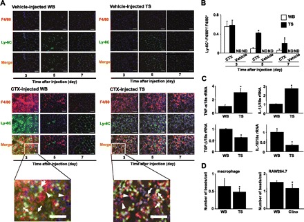
Skeletal muscle is one of the most sensitive tissues to mechanical loading, and unloading inhibits the regeneration potential of skeletal muscle after injury. This study was designed to elucidate the specific effects of unloading stress on the function of immunocytes during muscle regeneration after injury. We examined immunocyte infiltration and muscle regeneration in cardiotoxin (CTX)-injected soleus muscles of tail-suspended (TS) mice. In CTX-injected TS mice, the cross-sectional area of regenerating myofibers was smaller than that of weight-bearing (WB) mice, indicating that unloading delays muscle regeneration following CTX-induced skeletal muscle damage. Delayed infiltration of macrophages into the injured skeletal muscle was observed in CTX-injected TS mice. Neutrophils and macrophages in CTX-injected TS muscle were presented over a longer period at the injury sites compared with those in CTX-injected WB muscle. Disturbance of activation and differentiation of satellite cells was also observed in CTX-injected TS mice. Further analysis showed that the macrophages in soleus muscles were mainly Ly-6C-positive proinflammatory macrophages, with high expression of tumor necrosis factor-α and interleukin-1β, indicating that unloading causes preferential accumulation and persistence of proinflammatory macrophages in the injured muscle. The phagocytic and myotube formation properties of macrophages from CTX-injected TS skeletal muscle were suppressed compared with those from CTX-injected WB skeletal muscle. We concluded that the disturbed muscle regeneration under unloading is due to impaired macrophage function, inhibition of satellite cell activation, and their cooperation.


Figures

Fig. 1.
Fig. 1.
Histological analysis of muscle regeneration. A: representative sections (8-μm thickness) from soleus muscles of weight-bearing (WB) and tail suspension (TS) mice on day 14 after cardiotoxin (CTX) or vehicle injection were stained with hematoxylin and eosin (H and E). Arrows indicate the centralized nuclei. We observed 45 high-power fields per 15 sections in 5 mice/group in an experiment. Scale bar = 100 μm. B: calculated cross-sectional area (CSA) of myofibers in 45 high-power fields per 15 sections in 5 mice/group are expressed as means ± SD.
Fig. 2.
Fig. 2.
Time-dependent histological changes in soleus muscle after CTX injection. A: H and E-stained representative sections (8-μm thickness) of soleus muscles of WB and TS mice obtained at the indicated time points after injection of CTX or vehicle. Scale bar = 100 μm. B: the number of necrotic myofibers in 45 high-power fields per 15 sections in 5 mice per time point per group were counted and expressed as means ± SD. *P < 0.05 compared with vehicle injection, #P < 0.05 compared with CTX-injected WB mice.
Fig. 3.
Fig. 3.
Identification of infiltrated cells into injured muscles. A and C: representative sections (8-μm thickness) from soleus muscles of WB and TS mice obtained at the indicated time points after CTX or vehicle injection were immunostained with an anti-Gr-1 (green) antibody and Hoechst 33342 (blue) (A), or an anti-F4/80 (green) antibody and Hoechst 33342 (blue) (C). Scale bar = 100 μm. B and D: Gr-1-positive cells (B) and F4/80-positive cells (D) were counted in 45 high-power fields per 15 sections in 5 mice per time point per group and expressed as means ± SD. *P < 0.05 compared with vehicle injection, #P < 0.05 compared with CTX-injected WB mice.
Fig. 4.
Fig. 4.
Identification of proinflammatory and anti-inflammatory macrophages in injured muscles. A: representative sections (8 μm thickness) of soleus muscles of WB and TS mice obtained at the indicated time points after CTX or vehicle injection were immunostained with an anti-Ly-6C (green) antibody, anti-F4/80 (Red) antibody, and Hoechst 33342 (blue). We observed 45 high-power fields per 15 sections in 5 mice per time point per group in an experiment. Scale bar = 50 μm. B: we counted the number of yellow-colored cells (indicated by arrows in bottom panels) and red-colored cells (indicated by arrowheads in bottom panels) as Ly-6C-positive-F4/80 positive macrophages and Ly-6C-negative-F4/80 positive macrophages, respectively, in 45 high-power fields per 15 sections in 5 mice per time point per group. The ratio of yellow-colored cells to total macrophages (yellow-colored cells and red-colored cells) was calculated and is expressed as means ± SD. *P < 0.05 compared with CTX-injected WB mice. C: gene expression levels of proinflammatory and anti-inflammatory cytokines in macrophages isolated from muscles of WB and TS mice obtained on day 5 after CTX injection were assessed by real-time reverse transcription and polymerase chain reaction (RT-PCR). The fluorescence ratio of target gene cDNA to 18s ribosomal RNA (18s rRNA), a housekeeping gene, was calculated. Data are means ± SD of 3 mice. *P < 0.05 compared with CTX-injected WB mice. D: macrophages were isolated from muscles of WB and TS mice obtained on day 2 or 3 after CTX injection, respectively. RAW264.7 cells were cultured with or without clinorotation (clino) for 24 h. These cells were cultured with 2 μm fluoresbrite yellow green microspheres. One hour later, the number of microspheres in cells was counted in 12 high-power fields in 3 individual dishes and expressed as means ± SD. *P < 0.05 compared with WB conditions.
Fig. 5.
Fig. 5.
Stimulatory effects of macrophages on myotube formation. A: macrophages were prepared from soleus muscles of WB and TS mice on day 5 after CTX injection. Primary macrophages (9 × 10⁴ cells/well) were seeded on cultured primary myoblasts (3 × 10⁴ cells/well). These cells were cocultured for 3 days, and cells were immunostained with an anti-embryonic MyHC (green) and Hoechst 33342 (blue). Arrows indicate centrally located nuclei. Scale bar = 100 μm. B: myotube formation was estimated by counting the number of embryonic MyHC-positive multinuclear myotube in 12 high-power fields in 4 individual dishes. *P < 0.05 compared with myoblasts without macrophages, #P < 0.05 compared with myoblasts + macrophages from TS muscle.
Fig. 6.
Fig. 6.
Delayed activation of satellite cells in tail-suspended mice. A–D: representative sections (8 μm thickness) of soleus muscles of WB and TS mice were obtained at the indicated time points after injection of CTX or vehicle. A: representative sections were immunostained with an anti-MyoD (green) antibody and Hoechst 33342 (blue). Arrowheads: MyoD-positive mononuclear cells. Scale bar = 100 μm. B: MyoD-positive mononuclear cells were counted in 45 high-power fields per 15 sections in 5 mice per time point per group and are expressed as means ± SD. *P < 0.05 compared with vehicle injection, #P < 0.05 compared with CTX-injected WB mice. C: representative sections were immunostained with an anti-embryonic MyHC (green) and Hoechst 33342 (blue). Scale bar = 100 μm. D: embryonic MyHC-positive cells were counted in 45 high-power fields per 15 sections in 5 mice per time point per group and are expressed as means ± SD. *P < 0.05 compared with vehicle injection, #P < 0.05 compared with WB mice.
Fig. 7.
Fig. 7.
Kinetics of gene expression of MAFbx/Atrogin-1 and MuRF-1 in soleus muscles. A and B: levels of MAFbx/Atrogin-1 (A) and MuRF-1 (B) gene transcripts in soleus muscles of WB and TS mice on the indicated days after CTX or vehicle injection were assessed by real-time RT-PCR. The fluorescence ratio of target gene cDNA to glyceraldehyde-3-phosphate dehydrogenase (GAPDH), a housekeeping gene, was calculated. Data are means ± SD of 5 mice per time point per group. *P < 0.05 compared with vehicle injection.

Similar articles

Cited by

References

    1. Arnold L, Henry A, Poron F, Baba-Amer Y, van Rooijen N, Plonquet A, Gherardi RK, Chazaud B. Inflammatory monocytes recruited after skeletal muscle injury switch into antiinflammatory macrophages to support myogenesis. J Exp Med 204: 1057–1069, 2007 - PMC - PubMed
    1. Authier FJ, Chazaud B, Plonquet A, Eliezer-Vanerot MC, Poron F, Belec L, Barlovatz-Meimon G, Gherardi RK. Differential expression of the IL-1 system components during in vitro myogenesis: implication of IL-1β in induction of myogenic cell apoptosis. Cell Death Differ 6: 1012–1021, 1999 - PubMed
    1. Champion JA, Walker A, Mitragotri S. Role of particle size in phagocytosis of polymeric microspheres. Pharm Res 25: 1815–1821, 2008 - PMC - PubMed
    1. Charo IF, Taubman MB. Chemokines in the pathogenesis of vascular disease. Circ Res 95: 858–866, 2004 - PubMed
    1. Chazaud B, Sonnet C, Lafuste P, Bassez G, Rimaniol AC, Poron F, Authier FJ, Dreyfus PA, Gherardi RK. Satellite cells attract monocytes and use macrophages as a support to escape apoptosis and enhance muscle growth. J Cell Biol 163: 1133–1143, 2003 - PMC - PubMed

Publication types

MeSH terms

LinkOut - more resources