. 2012 Apr 20;287(17):13959-71.
doi: 10.1074/jbc.M111.288746. Epub 2012 Mar 1.

Low-density lipoprotein receptor represents an apolipoprotein E-independent pathway of Aβ uptake and degradation by astrocytes

Affiliations

Low-density lipoprotein receptor represents an apolipoprotein E-independent pathway of Aβ uptake and degradation by astrocytes

Jacob M Basak et al. J Biol Chem. .

Abstract

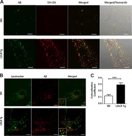
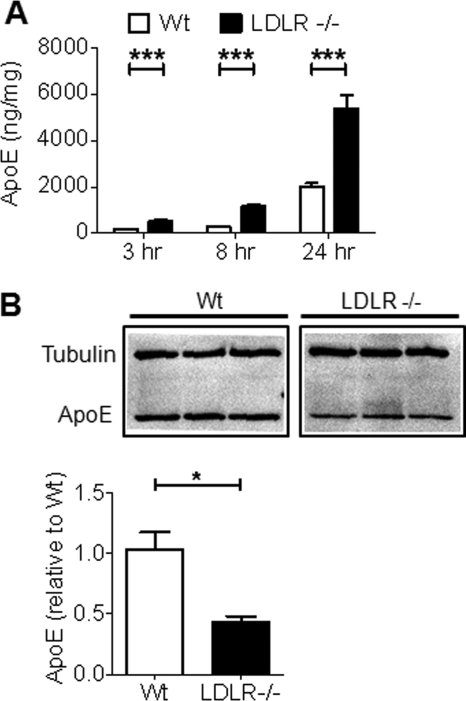
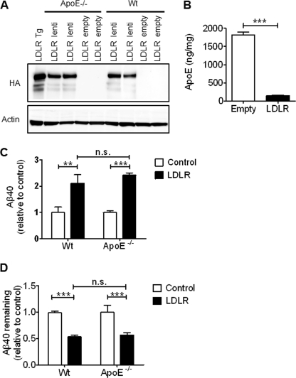
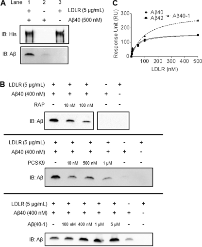
Accumulation of the amyloid β (Aβ) peptide within the brain is hypothesized to be one of the main causes underlying the pathogenic events that occur in Alzheimer disease (AD). Consequently, identifying pathways by which Aβ is cleared from the brain is crucial for better understanding of the disease pathogenesis and developing novel therapeutics. Cellular uptake and degradation by glial cells is one means by which Aβ may be cleared from the brain. In the current study, we demonstrate that modulating levels of the low-density lipoprotein receptor (LDLR), a cell surface receptor that regulates the amount of apolipoprotein E (apoE) in the brain, altered both the uptake and degradation of Aβ by astrocytes. Deletion of LDLR caused a decrease in Aβ uptake, whereas increasing LDLR levels significantly enhanced both the uptake and clearance of Aβ. Increasing LDLR levels also enhanced the cellular degradation of Aβ and facilitated the vesicular transport of Aβ to lysosomes. Despite the fact that LDLR regulated the uptake of apoE by astrocytes, we found that the effect of LDLR on Aβ uptake and clearance occurred in the absence of apoE. Finally, we provide evidence that Aβ can directly bind to LDLR, suggesting that an interaction between LDLR and Aβ could be responsible for LDLR-mediated Aβ uptake. Therefore, these results identify LDLR as a receptor that mediates Aβ uptake and clearance by astrocytes, and provide evidence that increasing glial LDLR levels may promote Aβ degradation within the brain.


Figures

FIGURE 1.
FIGURE 1.
Increased LDLR levels alter the extracellular and intracellular levels of apoE in primary astrocytes. Primary astrocytes were cultured from the cortices of both WT and LDLR transgenic mice. The LDLR transgene is expressed under control of the mouse prion promoter and also contains a hemagglutinin (HA) tag. A, LDLR and HA levels in the cells were measured by immunoblot. Unglycosylated LDLR migrates at 90 kDa and several glycosylated species of the protein migrate between 100 and 150 kDa. Representative images are shown. B, the functional effect of increased LDLR levels on apoE uptake was assessed by measuring the levels of endogenously produced apoE in the culture media. Primary astrocytes were incubated for the indicated time points in serum-free medium and the amount of apoE was measured by ELISA. Mean ± S.E. (n = 4), * denotes p < 0.05, ** denotes p < 0.01, *** denotes p < 0.001. C, the amount of cell-associated apoE was also measured by immunoblot of the cell lysates obtained after a 24-h incubation. A representative image is shown.
FIGURE 2.
FIGURE 2.
LDLR overexpression enhances the uptake and clearance of Aβ by primary astrocytes. Primary astrocytes from either WT or LDLR transgenic mice were incubated with soluble (A) Aβ40 or (B) Aβ42 (2 μg/ml) for 3 h at 37 °C. The cells were then washed with PBS, incubated with trypsin to remove cell surface bound Aβ, and lysed in Triton X-100 lysis buffer. The cell-internalized Aβ was then assessed by ELISA. Mean ± S.E. (n ≥ 4), *** denotes p < 0.001. C, immunoblot analysis for Aβ was also performed on the cell lysates. Representative images are shown. Aβ clearance was assessed by the addition of either (D) Aβ40 or (E) Aβ42 (2 μg/ml) to the media of primary astrocytes. After 24 h, the levels of Aβ remaining in the medium along with the starting amount of Aβ were measured by ELISA. Mean ± S.E. (n ≥ 4), * denotes p < 0.05, *** denotes p < 0.001.
FIGURE 3.
FIGURE 3.
LDLR overexpression increases the cellular degradation of Aβ by primary astrocytes. A, schematic diagram of the experiments used to measure degradation of 125I-Aβ by primary astrocytes. 125I-Aβ was added to primary astrocytes from either WT or LDLR Tg mice at the indicated time points. After each time point, media was collected and a TCA precipitation was performed to detect degraded Aβ. B, the supernatant (sup) and pellet counts/min are plotted as a function of time. Representative data from one experiment is shown. Experiment was repeated three times with similar results. C, degraded Aβ was quantified by calculating the percent of Aβ degraded as a percent of the total intact Aβ added. Mean ± S.E., * denotes p < 0.05, ** denotes p < 0.01. D, to measure the ability of astrocyte-conditioned media to degrade Aβ, media was collected from either WT or LDLR Tg primary astrocytes. 125I-Aβ was then added to the astrocyte-conditioned medium at the indicated time points and a TCA precipitation was performed. The supernatant (sup) and pellet counts/min are plotted as a function of time. Representative data from one experiment is shown. The experiment was repeated two times with similar results. E, degraded Aβ was quantified by calculating the percent of Aβ degraded as a percent of the total intact Aβ added. Mean ± S.E., * denotes p < 0.05, n.s., not significant.
FIGURE 4.
FIGURE 4.
LDLR facilitates Aβ trafficking to lysosomes through a similar pathway as lipoprotein particles. A, to demonstrate that increasing LDLR levels promotes the transport of Aβ in similar vesicles as lipoprotein particles, WT and LDLR Tg primary astrocytes were incubated with fluorescent Aβ42 (3 μg/ml) and DiI-LDL (0.5 μg/ml) for 3 h at 37 °C. The cells were then washed and imaged using confocal microscopy. Overlap of Aβ and the DiI-LDL signal was observed in the LDLR Tg cells. B, to observe Aβ uptake into lysosomal compartments, WT and LDLR Tg primary astrocytes were incubated with fluorescent Aβ42 (2 μg/ml) for 3 h at 37 °C. The cells were then washed and 50 nm LysoTracker was added to the cells for 15 min. The cells were then washed again and imaged using confocal microscopy. C, colocalization of the Aβ and LysoTracker signal was analyzed and quantified. Mean ± S.E., *** denotes p < 0.001. Error bar represents 10 μm.
FIGURE 5.
FIGURE 5.
Deletion of LDLR alters the extracellular and intracellular levels of apoE. Primary astrocytes were cultured from the cortices of WT and LDLR−/− mice. A, to show that LDLR deletion alters lipoprotein levels in astrocytes, apoE uptake was assessed by measuring the levels of endogenously produced apoE in the culture media. Primary astrocytes were incubated for the indicated time points in serum-free medium and the amount of apoE in the medium was measured by ELISA. Mean ± S.E. (n ≥ 4). *** denotes p < 0.001. B, the amount of cell-associated apoE was also measured by immunoblot of the cell lysates obtained after the 24-h incubation. Quantification of the apoE band intensity normalized to tubulin intensity is shown below the image.
FIGURE 6.
FIGURE 6.
Lack of LDLR impairs Aβ uptake in astrocytes. To assess the effect of LDLR deletion on Aβ uptake, WT and LDLR−/− astrocytes were incubated with Aβ40 (2 μg/ml) for 3 h. The cells were then washed with PBS, incubated with trypsin to remove cell surface-bound Aβ, and lysed in Triton X-100 lysis buffer. The amount of Aβ in the cell lysate was then assessed by ELISA (A) and immunoblot (B). For the immunoblot, a representative image is shown. Mean ± S.E. (n ≥ 4). *** denotes p < 0.001. C, to verify the effect of LDLR deletion on Aβ uptake, LDLR function was restored in the LDLR−/− astrocytes by transduction with an LDLR lentivirus. Aβ uptake was then assessed as in A and compared with the level of uptake by LDLR−/− cells transduced with control lentivirus and WT cells. Mean ± S.E. (n ≥ 4). ** denotes p < 0.01. D, the effect of LDLR deletion on Aβ clearance was assessed by the addition of Aβ40 (2 μg/ml) to the WT and LDLR−/− astrocytes media. After 24 h, the amount of Aβ remaining was measured by ELISA and compared with the starting amount. Mean ± S.E. (n ≥ 4). * denotes p < 0.05; n.s., not significant.
FIGURE 7.
FIGURE 7.
The effect of LDLR on Aβ uptake and clearance is not dependent on apoE. A, to determine whether the effect of LDLR on Aβ uptake and clearance requires the presence of apoE, LDLR was expressed in apoE−/− and WT primary astrocytes via lentiviral transduction. LDLR expression was confirmed by immunoblot for HA. LDLR Tg astrocyte lysate is shown for comparison. B, to confirm that the LDLR protein expressed after lentiviral transduction was functional, WT cells were transduced and the amount of endogenously produced apoE was measured by ELISA in the cell medium after a 24-h incubation. Mean ± S.E. (n ≥ 4). ** denotes p < 0.01. C, Aβ uptake was measured in WT and apoE−/− primary astrocytes transduced with LDLR lentivirus. Aβ40 (2 μg/ml) was incubated with the cells for 3 h. The cells were then washed with PBS, treated with trypsin to remove cell surface-bound Aβ, and lysed in Triton X-100 lysis buffer. The cell-internalized Aβ was then measured by ELISA. Control samples were transduced with the empty lentivirus. Mean ± S.E. (n ≥ 4). ** denotes p < 0.01; *** denotes p < 0.001; n.s., not significant. D, Aβ clearance was assessed by the addition of Aβ40 (2 μg/ml) to the media of WT and ApoE−/− astrocytes transduced with the LDLR lentivirus. After 24 h, the amount of Aβ remaining was measured by ELISA and compared with cells transduced with empty lentivirus. Mean ± S.E. (n ≥ 4). *** denotes p < 0.001; n.s., not significant.
FIGURE 8.
FIGURE 8.
Direct interaction between Aβ and LDLR. A, to assess whether Aβ could directly associate with LDLR, Aβ40 (500 nm) and recombinant extracellular LDLR (5 μg/ml) were incubated together and immunoprecipitated using anti-His beads to pull down LDLR. The isolated proteins were then eluted from the beads and subjected to SDS-PAGE and immunoblot analysis for LDLR (His) and Aβ. Control experiments included incubating Aβ40 alone with anti-His beads and immunoprecipitating LDLR without the addition of Aβ40. B, the specificity of the binding of Aβ to LDLR was determined by performing competition experiments with known LDLR ligands. Increasing amounts of either RAP or PCSK9 were preincubated with recombinant extracellular LDLR for 2 h, and Aβ40 was then added to the protein mixture and incubated at 37 °C for 4 h. LDLR was then immunoprecipitated using anti-His beads, and the eluted samples were subjected to SDS-PAGE and immunoblot analysis for Aβ. The experiment was also repeated using Aβ(40-1) as a competing peptide. C, surface plasmon resonance was used to measure the interaction between the extracellular domain of LDLR and Aβ. Aβ40, Aβ42, or Aβ(40-1) were immobilized on the SPR chip and various concentrations of LDLR were flown over the surface. To calculate the dissociation constant for the interaction (KD), we plotted the resonance units as a function of LDLR concentration.

Similar articles

Cited by

References

    1. Holtzman D. M., Morris J. C., Goate A. M. (2011) Alzheimer disease, the challenge of the second century. Sci. Transl. Med. 3, 77sr1. - PMC - PubMed
    1. Hardy J., Selkoe D. J. (2002) The amyloid hypothesis of Alzheimer disease. Progress and problems on the road to therapeutics. Science 297, 353–356 - PubMed
    1. De Strooper B. (2010) Proteases and proteolysis in Alzheimer disease. A multifactorial view on the disease process. Physiol. Rev. 90, 465–494 - PubMed
    1. Hardy J. (2006) A hundred years of Alzheimer disease research. Neuron 52, 3–13 - PubMed
    1. Selkoe D. J. (2001) Clearing the brain's amyloid cobwebs. Neuron 32, 177–180 - PubMed

Publication types