. 2012 Apr 13;287(16):13228-38.
doi: 10.1074/jbc.M111.334524. Epub 2012 Mar 1.

Characterization of EHop-016, novel small molecule inhibitor of Rac GTPase

Affiliations

Characterization of EHop-016, novel small molecule inhibitor of Rac GTPase

Brenda L Montalvo-Ortiz et al. J Biol Chem. .

Abstract

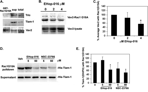
The Rho GTPase Rac regulates actin cytoskeleton reorganization to form cell surface extensions (lamellipodia) required for cell migration/invasion during cancer metastasis. Rac hyperactivation and overexpression are associated with aggressive cancers; thus, interference of the interaction of Rac with its direct upstream activators, guanine nucleotide exchange factors (GEFs), is a viable strategy for inhibiting Rac activity. We synthesized EHop-016, a novel inhibitor of Rac activity, based on the structure of the established Rac/Rac GEF inhibitor NSC23766. Herein, we demonstrate that EHop-016 inhibits Rac activity in the MDA-MB-435 metastatic cancer cells that overexpress Rac and exhibits high endogenous Rac activity. The IC(50) of 1.1 μM for Rac inhibition by EHop-016 is ∼100-fold lower than for NSC23766. EHop-016 is specific for Rac1 and Rac3 at concentrations of ≤5 μM. At higher concentrations, EHop-016 inhibits the close homolog Cdc42. In MDA-MB-435 cells that demonstrate high active levels of the Rac GEF Vav2, EHop-016 inhibits the association of Vav2 with a nucleotide-free Rac1(G15A), which has a high affinity for activated GEFs. EHop-016 also inhibits the Rac activity of MDA-MB-231 metastatic breast cancer cells and reduces Rac-directed lamellipodia formation in both cell lines. EHop-016 decreases Rac downstream effects of PAK1 (p21-activated kinase 1) activity and directed migration of metastatic cancer cells. Moreover, at effective concentrations (<5 μM), EHop-016 does not affect the viability of transformed mammary epithelial cells (MCF-10A) and reduces viability of MDA-MB-435 cells by only 20%. Therefore, EHop-016 holds promise as a targeted therapeutic agent for the treatment of metastatic cancers with high Rac activity.


Figures

FIGURE 1.
FIGURE 1.
Synthesis and docking of EHop-016 into the putative GEF binding pocket of Rac1. A, synthetic scheme for the preparation of EHop-016. The synthesis was performed by a two-step approach as described (57). B, EHop-016 docked into the GEF binding pocket of Rac1 and its comparison with the position of NSC23766 in the crystal structure of the Rac1-NSC23766 complex.
FIGURE 2.
FIGURE 2.
Effect of EHop-016 and NSC23766 on Rac activity. MDA-MB-435 cells were treated with vehicle (0.1% DMSO) or varying concentrations of EHop-016 (0–10 μm) or NSC23766 (0–100 μm) for 24 h. Cell lysates were subjected to the G-LISA Rac1 activation assay (Cytoskeleton, Inc.). IC50 curves for percentage Rac activity are relative to vehicle from three biological replicates each with two technical replicates. Error bars, S.D. Four-parameter dose-response curves generated using GraphPad Prism® are shown.
FIGURE 3.
FIGURE 3.
Effect of EHop-016 on Rho GTPase activity. A and B, MDA-MB-435 cells were treated with vehicle (0) or EHop-016 at the indicated concentrations (1, 2, 4, 5, or 10 μm) for 24 h. Cell lysates were subjected to a pull-down assay using a GST-CRIB domain of PAK and Western blotted for Rac3 or Cdc42. C, a GST-RBD domain of rhotekin was used to pull down Rho-GTP and detected by Western blotting with anti-RhoA antibody. The average percentage of Rho GTPase activity was calculated from the integrated density of positive bands of Rho GTPase-GTP from a pull-down/total Rho GTPase in cell lysate for each treatment and each Rho GTPase (Rac3, Cdc42, or RhoA) relative to vehicle controls. A, representative Western blot of pull-downs immunostained for Rac3-GTP (top row) or total Rac3 in cell lysate (bottom row) (n = 3). B, representative Western blots (from the same experiment) of pull-downs immunostained for Cdc42-GTP (top row) or total Cdc42 in cell lysate (bottom row) (n = 2). C, representative Western blots of pull-downs immunostained for RhoA-GTP (top row) or total RhoA in cell lysate (bottom row), n = 3. Western blots show positive bands for Rho GTPases at ∼21 kDa.
FIGURE 4.
FIGURE 4.
Effect of EHop-016 on Rac/Rac GEF interaction. MDA-MB-435 cells were lysed and incubated with glutathione-agarose-coupled GST-Rac1(G15A) beads to pull down activated Rac GEFs. A, a representative Western blot (from the same experiment) of GST-Rac1(G15A) pull-down, supernatant (sup) from the incubation, and cell lysates, immunostained for Trio, Tiam-1, or Vav2. B, GST-Rac1(G15A) beads were preincubated with vehicle (0), or 2 or 4 μm EHop-016 prior to incubation with MDA-MB-435 cell lysates. A representative Western blot (from the same experiment) (n = 3) immunostained for Vav2 is shown. Top row, pull-down; bottom row, total cell lysate. C, quantification of the percentage of average Vav2 (two bands at ∼100 kDa) associated with the Rac1(G15A) beads from pull-down assays in the presence or absence of EHop-016. ImageJ software was used to quantify the integrated density of positive bands (∼100 kDa) from Vav2 Western blots of pull-downs. Data are represented relative to vehicle controls (100%). D and E, purified His-tagged Tiam-1 DH/PH domain was added at a concentration of 2:1 to Rac1(G15A) beads that were preincubated with vehicle (Veh) or Ehop-016 or NSC23766 at the indicated concentrations for 1 h at 4 °C. Pull-downs were washed and immunoblotted with an anti-His antibody to detect His-Tiam-1 as a ∼45 kDa band. D, a representative Western blot (from the same experiment). Top, GST-Rac1(G15A) pull-downs; bottom, supernatants. E, quantification of percentage of Tiam-1 (DH/PH) domain associated with Rac1(G15A) beads in the presence or absence of EHop-016 or NSC23766. n = 3. Error bars, S.D. *, statistical significance compared with vehicle controls (p ≤ 0.05).
FIGURE 5.
FIGURE 5.
Effect of EHop-016 on Rac activity and the actin cytoskeleton of metastatic cancer cells. A, MDA-MB-231 or MDA-MB-435 cells were treated with vehicle (0) or EHop-016 at the indicated concentrations (2 or 4 μm) for 24 h. Cell lysates were subjected to a pull-down assay using a GST-CRIB domain of PAK and Western blotted for Rac (Rac1, -2, and -3). Representative Western blot (from the same experiment) of pull-downs immunostained for Rac-GTP (top row) or total Rac in cell lysate (bottom row). n = 3. B, MDA-MB-231 or MDA-MB-435 metastatic breast cancer cells were treated with vehicle or EHop-016 at 2 or 4 μm for 24 h to determine changes in actin cytoskeletal structures. Cells were fixed, permeabilized, and stained with rhodamine phalloidin to visualize F-actin. Top, representative micrographs are shown at ×600 magnification. Arrows, lamellipodia; arrowheads, filopodia. Bottom, percentage of cells that demonstrated lamellipodia was quantified for each treatment from 10 representative microscopic fields. Error bars, S.E. *, statistical significance compared with vehicle controls (p ≤ 0.001).
FIGURE 6.
FIGURE 6.
Effect of EHop-016 on Rac-regulated PAK activity and directed cell migration. A, MDA-MB-435 cells were treated with vehicle (0) or 2 or 4 μm EHop-016 for 24 h, and the cells were lysed and Western blotted for active phospho-PAKThr-423 (p-PAK; upper band) or total PAK (lower band). B, quantification of positive bands from Western blots (65 kDa) using ImageJ software. The integrated density of the phospho-PAK band was divided by the integrated density of total PAK band from the same sample. EHop-016 treatments are presented relative to vehicle controls (100%). n = 3. Error bars, S.D. *, statistical significance compared with vehicle controls (p ≤ 0.05). C, MDA-MB-435 cells treated with vehicle (0) or EHop-016 (1–5 μm) for 24 h were subjected to a Transwell migration assay. The number of cells that migrated to the underside of the top well in response to serum in the bottom well was quantified for each treatment. Top, representative micrographs of propidium iodide-stained cells for each treatment at ×200 magnification. Bottom, percentage of cells that migrated to the underside of a membrane with 8-μm diameter pores, relative to vehicle (100%). Results are shown for three biological replicates with two technical replicates per experiment. Error bars, S.E. *, statistical significance compared with vehicle controls (p ≤ 0.05).
FIGURE 7.
FIGURE 7.
Effect of EHop-016 on cell viability. Cell viability of MDA-MB-231, MDA-MB-435, or MCF-10A cells was measured using the 3-(4,5-dimethylthiazol-2-yl)-2,5-diphenyltetrazolium bromide cell survival and proliferation kit (Millipore, Inc.). The mean values ± S.E. (error bars) (n = 3) are presented relative to vehicle (100%). *, statistical significance compared with vehicle controls (p ≤ 0.05).

Similar articles

Cited by

References

    1. Ridley A. J. (2006) Rho GTPases and actin dynamics in membrane protrusions and vesicle trafficking. Trends Cell Biol. 16, 522–529 - PubMed
    1. Mack N. A., Whalley H. J., Castillo-Lluva S., Malliri A. (2011) The diverse roles of Rac signaling in tumorigenesis. Cell Cycle 10, 1571–1581 - PMC - PubMed
    1. Lazer G., Katzav S. (2011) Guanine nucleotide exchange factors for RhoGTPases. Good therapeutic targets for cancer therapy? Cell. Signal. 23, 969–979 - PubMed
    1. Parri M., Chiarugi P. (2010) Rac and Rho GTPases in cancer cell motility control. Cell Commun. Signal. 8, 23. - PMC - PubMed
    1. Mulloy J. C., Cancelas J. A., Filippi M. D., Kalfa T. A., Guo F., Zheng Y. (2010) Rho GTPases in hematopoiesis and hemopathies. Blood 115, 936–947 - PMC - PubMed

Publication types

MeSH terms