. 2012 Aug;18(8):417-24.
doi: 10.1093/molehr/gas013. Epub 2012 Feb 29.

The expression profile of C19MC microRNAs in primary human trophoblast cells and exosomes

Affiliations

The expression profile of C19MC microRNAs in primary human trophoblast cells and exosomes

R B Donker et al. Mol Hum Reprod. 2012 Aug.

Abstract

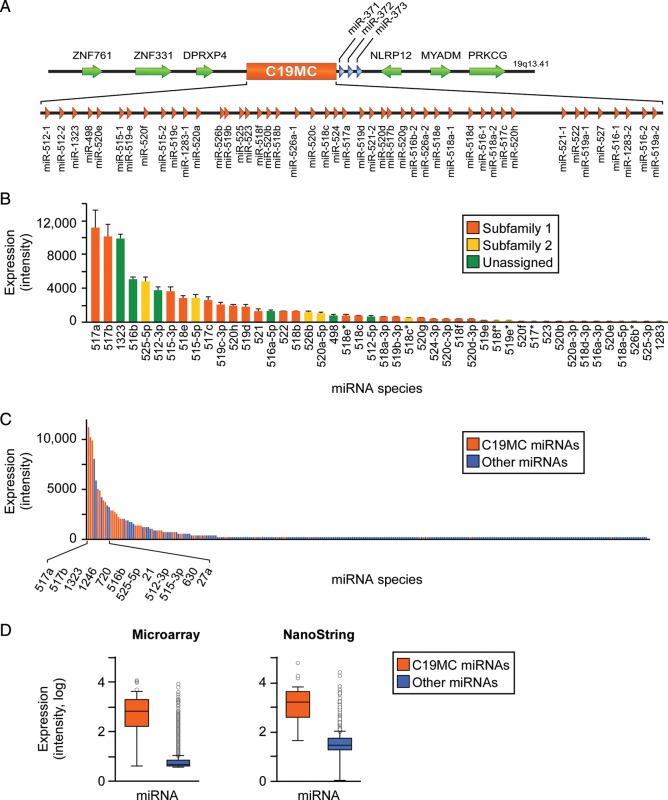
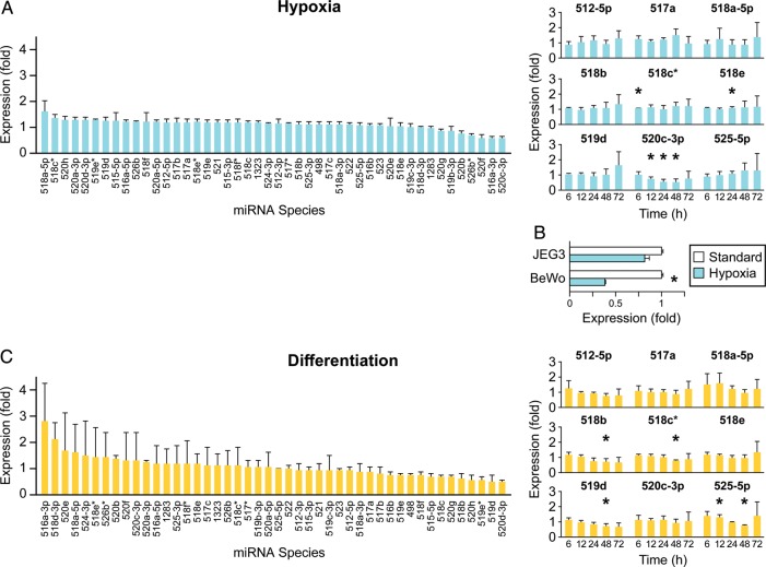
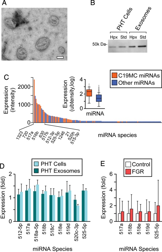
The largest gene cluster of human microRNAs (miRNAs), the chromosome 19 miRNA cluster (C19MC), is exclusively expressed in the placenta and in undifferentiated cells. The precise expression pattern and function of C19MC members are unknown. We sought to profile the relative expression of C19MC miRNAs in primary human trophoblast (PHT) cells and exosomes. Using high-throughput profiling, confirmed by PCR, we found that C19MC miRNAs are among the most abundant miRNAs in term human trophoblasts. Hypoxic stress selectively reduced miR-520c-3p expression at certain time-points with no effect on other C19MC miRNAs. Similarly, differentiation in vitro had a negligible effect on C19MC miRNAs. We found that C19MC miRNAs are the predominant miRNA species expressed in exosomes released from PHT, resembling the profile of trophoblastic cellular miRNA. Predictably, we detected the similar levels of circulating C19MC miRNAs in the serum of healthy pregnant women at term and in women with pregnancies complicated by fetal growth restriction. Our data define the relative expression levels of C19MC miRNAs in trophoblasts and exosomes, and suggest that C19MC miRNAs function in placental-maternal signaling.


Figures

Figure 1
Figure 1
C19MC miRNAs landscape in term PHT cells. (A) Genomic organization of the C19MC miRNA cluster. (B) The expression of C19MC miRNAs species in PHT cells, as determined by miRNA microarrays (t= 48 h, n= 4). The expression represents the mean of normalized signal intensities, which are stratified by miRNA family as described in the text. (C) C19MC miRNAs comprise a major part of PHT miRNAs, as determined by miRNA microarrays (t = 48 h, n = 4). The expression represents the mean of normalized signal intensities. (D) The expression level of all C19MC miRNAs versus all other miRNAs in PHT cells, as measured by microarray or NanoString (t = 48 h, n = 4 for both analyses). Boxplots represent mean, 25th and 75th percentile, and outliers (differences were significant at P < 0.05). All error bars represent SD.
Figure 2
Figure 2
The effect of hypoxia or in vitro differentiation on the expression of C19MC miRNAs. (A) Hypoxia does not affect C19MC miRNA expression, except for down-regulation of miR-520c-3p at 12, 24 and 48 h of exposure to hypoxia, as determined by high-throughput microarray profiling (left panel, t = 48 h, n = 4) or RT-qPCR (right panel, t = 48 h, n = 4). Fold expression represents mean of fold change in hypoxia (O2 <1%) over standard conditions (O2= 21%). (B) The expression of miR-520c-3p in placental cell lines after 36–48 h of exposure to hypoxia, as determined by RT-qPCR (representative experiment of n = 2 for BeWo, and n= 3 for JEG-3 cells). Fold expression represents mean of fold change in hypoxia (O2 <1%) over control (O2= 21%). *Denotes P < 0.05. (C) C19MC miRNA expression is not affected by PHT differentiation in vitro, as determined by high-throughput microarray profiling (left panel, t = 48 h, n = 4) or RT-qPCR (right panel, t = 48 h, n = 4). Fold expression represents the mean of fold change at t = 48 h over t = 0 h. *Denotes P < 0.05. All error bars represent SD.
Figure 3
Figure 3
The profile of C19MC miRNAs in PHT exosomes resembles that of PHT cells. (A) PHT cells release exosomes and microvesicles of diverse sizes (see ‘Materials and Methods’ section for details of electron microscopy measurements). Bar = 100 nm. (B) PHT microvesicle population is enriched for the exosome marker TSG101, in standard (SD) and hypoxic conditions. Expression determined by western immunoblotting. (C) C19MC miRNAs comprise the majority of miRNAs in PHT exosomes, as determined by miRNA microarray without replicates (t = 48 h). Expression represents signal intensity. (D) The relative expression level of C19MC miRNA in PHT exosomes resembles the expression level detected in PHT cells cultured in hypoxic conditions, as determined by RT-qPCR (t = 48 h, n = 3). The fold expression represents mean fold change in hypoxia (O2 <1%) over normoxia (O2= 21%). (E) Circulating C19MC miRNA species are detected in the plasma of normal human pregnancy or pregnancy complicated by FGR as described in ‘Materials and Methods’ section. MiRNAs were detected using RT-qPCR (n = 14 for each group), with fold expression in normal pregnancy defined as 1. All error bars represent SD.

Similar articles

Cited by

References

    1. Arroyo JD, Chevillet JR, Kroh EM, Ruf IK, Pritchard CC, Gibson DF, Mitchell PS, Bennett CF, Pogosova-Agadjanyan EL, Stirewalt DL, et al. Argonaute2 complexes carry a population of circulating microRNAs independent of vesicles in human plasma. Proc Natl Acad Sci USA. 2011;108:5003–5008. - PMC - PubMed
    1. Atay S, Gercel-Taylor C, Kesimer M, Taylor DD. Morphologic and proteomic characterization of exosomes released by cultured extravillous trophoblast cells. Exp Cell Res. 2011;317:1192–1202. - PubMed
    1. Barad O, Meiri E, Avniel A, Aharonov R, Barzilai A, Bentwich I, Einav U, Gilad S, Hurban P, Karov Y, et al. MicroRNA expression detected by oligonucleotide microarrays: system establishment and expression profiling in human tissues. Genome Res. 2004;14:2486–2494. - PMC - PubMed
    1. Bentwich I, Avniel A, Karov Y, Aharonov R, Gilad S, Barad O, Barzilai A, Einat P, Einav U, Meiri E, et al. Identification of hundreds of conserved and nonconserved human microRNAs. Nat Genet. 2005;37:766–770. - PubMed
    1. Bortolin-Cavaille ML, Dance M, Weber M, Cavaille J. C19MC microRNAs are processed from introns of large Pol-II, non-protein-coding transcripts. Nucleic Acids Res. 2009;37:3464–3473. - PMC - PubMed

Publication types