. 2012 Mar 20;109(12):4639-44.
doi: 10.1073/pnas.1116269109. Epub 2012 Mar 1.

Identification of a cis-acting element that localizes mRNA to synapses

Affiliations

Identification of a cis-acting element that localizes mRNA to synapses

Elliott J Meer et al. Proc Natl Acad Sci U S A. .

Abstract

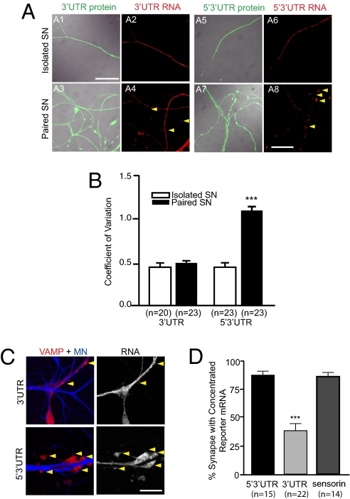
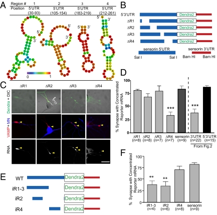
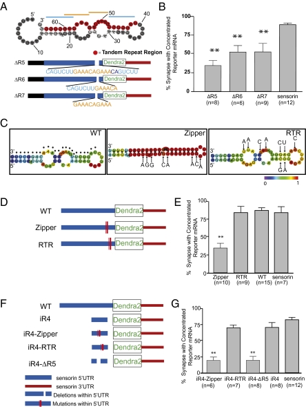
Messenger RNA (mRNA) localization and regulated translation can spatially restrict gene expression to each of the thousands of synaptic compartments formed by a single neuron. Although cis-acting RNA elements have been shown to direct localization of mRNAs from the soma into neuronal processes, less is known about signals that target transcripts specifically to synapses. In Aplysia sensory-motor neuronal cultures, synapse formation rapidly redistributes the mRNA encoding the peptide neurotransmitter sensorin from neuritic shafts into synapses. We find that the export of sensorin mRNA from soma to neurite and the localization to synapse are controlled by distinct signals. The 3' UTR is sufficient for export into distal neurites, whereas the 5' UTR is required for concentration of reporter mRNA at synapses. We have identified a 66-nt element in the 5' UTR of sensorin that is necessary and sufficient for synaptic mRNA localization. Mutational and chemical probing analyses are consistent with a role for secondary structure in this process.


Conflict of interest statement

The authors declare no conflict of interest.

Figures

Fig. 1.
Fig. 1.
The 3′ UTR of sensorin is sufficient to target reporter mRNA into distal neurites. pNEX vectors encoding translational reporters were microinjected into isolated Aplysia SNs in culture (DIV 2). Neurons were fixed (DIV 4) and processed for FISH with dendra antisense riboprobes. (A) Cartoons of reporter constructs in pNEX3 expression vector. (B) Representative images of reporter (dendra2) mRNA FISH in isolated SNs. (B1–B6) Neurolucida tracing of each SN; (B1′–B6′) FISH (detected with dendra antisense riboprobes for B1′–B5′ and with sensorin antisense riboprobes for B6′). The FISH signal only extends to distal neurites when the 3′ UTR of sensorin is present; distal localization is enhanced by 5′ UTR. (Scale bar, 100 μm.) (C) Quantification of the distribution of reporter mRNA within sensory neurites. Neurites were linearized and divided into proximal, middle, and distal segments. The percentage of total FISH signal in distal segments is shown (see also Fig. S1). ***P < 0.0001, Kruskal-Wallis one-way analysis of variance followed by Dunnett's multiple comparison test.
Fig. 2.
Fig. 2.
The 5′ UTR of sensorin is required for localizing reporter mRNA to synapse. Expression vectors encoding dendra2 reporters with either the 3′ UTR or both the 5′ and 3′ UTRs (5′3′ UTR) of sensorin were microinjected into Aplysia SN (DIV 2) cultured in isolation (isolated SN) or with MNs. Neurons were fixed (DIV 4) and processed for FISH using dendra2 antisense riboprobes. (A) Representative photomicrographs of dendra reporter protein (green, A1, A3, A5, A7) and mRNA (red, A2, A4, A6, A8) distribution in isolated and paired SNs. (Scale bar, 50 μm.) (B) Quantification of the change in distribution of reporter mRNA by measuring the CV (SD/mean) of the FISH signal. ***P < 0.0001, unpaired Student t test. (C) The 5′3′ UTR reporter or the 3′ UTR reporter was coexpressed with the presynaptic marker VAMP-mCherry in Aplysia SNs paired with target MNs on DIV 2. On DIV 4, the MN was injected with the volume filling dye Alexa Fluor 647 (blue), and images of VAMP-mCherry and blue Alexa Fluor were acquired, followed by fixation and FISH with dendra antisense riboprobes. Left: Merged VAMP/MN images of a coculture with SN overexpressing 5′3′ UTR or 3′ UTR reporter and VAMP-mCherry (VAMP-mCherry in red, MN in blue); Right: FISH signal for reporter mRNA. (Scale bar, 100 μm.) (D) Quantification of the percentage of synapses (VAMP-mCherry clusters adjacent to MN) containing reporter mRNA (error bars, SEM). ***P < 0.0001, one-way ANOVA followed by Dunnett's multiple comparison test. See also ref. and Fig. S2.
Fig. 3.
Fig. 3.
A region directly upstream of the sensorin translation start site, in the 5′ UTR, is necessary and sufficient for synaptic localization of the reporter mRNA. (A) Representative graphic depictions of potential stem-loop structures in the 5′ UTR of sensorin predicted by RNAfold (35); color scale denotes base pair probabilities. (B) Cartoon representation of deletions made to the reporter construct. (C) Representative images of deletion constructs ΔR1–4 expressed in SNs synaptically connected with MNs. Top: Photomicrograph of Dendra protein (SN, green) merged with DIC image. Middle: Synapses marked as VAMP-mCherry clusters (red) contacting the MN (blue, Alexa 647). Bottom: FISH images showing clustering of reporter mRNA. (D) Quantification of synaptic localization of reporter mRNAs as the percentage of synapses (VAMP-mCherry clusters adjacent to MN) containing reporter mRNA. (E) Cartoon of insertion constructs (iR1–4) in which the designated regions of the sensorin 5′ UTR were inserted into a control pNEX 5′ UTR to test their ability to localize the reporter mRNA to synapses. The mutants were coexpressed in SNs paired with target MNs at DIV 2, and neurons were fixed on DIV 4 and processed for FISH. (F) Percentage of synapses (VAMP-mCherry clusters adjacent to MN) containing reporter mRNA.
Fig. 4.
Fig. 4.
Stem-loop structure (66 nt) localizes reporter mRNA to synapses. (A) Predicted secondary structure of the region corresponding to R4 [RNAfold (35); highlighted in red is the 24-nt primary sequence containing a double-tandem 7mer-repeat (indicated with blue/orange lines)]. Mutants were generated in which the entire 24-nt (ΔR5), the first 13-nt set of repeat elements (ΔR6), or 10 nt of the center repeat (ΔR7) were deleted from the 5′3′ UTR reporter construct. (B) Mutants were coexpressed with VAMP-mCherry in SNs paired with target MNs, and the percentage of synapses containing reporter RNA was measured. **P < 0.01, one-way ANOVA followed by Dunnett's multiple comparison test compared with 3′ UTR reporter or endogenous sensorin. See also Figs. S2, S3, and S5. (C) Predicted secondary structures [RNAfold (35)] of WT (dots denote tandem repeat region described in Fig. 3); zipper construct, 8-point mutations were introduced to collapse the predicted secondary structure without disrupting the tandem repeat sequence; RTR construct, 9 mutations were introduced to disrupt the primary sequence of the tandem repeat region, while retaining predicted secondary structure. (D) Cartoon showing the location within the sensorin 5′ UTR of the mutations in the zipper and RTR constructs. The mutants were coexpressed in SNs paired with target MNs at DIV 2, and neurons were fixed on DIV 4 and processed for FISH. (E) Percentage of synapses (VAMP-mCherry clusters adjacent to MN) containing reporter mRNA. (F) Cartoon of mutant insertion reporter constructs, including WT iR4 (66 nt), iR4-RTR (iR4 with mutations shown in RTR in C), iR4-Zipper (iR4 with mutations shown in zipper in C), or iR4-ΔR5 (iR4 lacking the 24-nt repeat element). The mutants were coexpressed in SNs paired with target MNs at DIV 2, and neurons were fixed on DIV 4 and processed for FISH. (G) Percentage of synapses (VAMP-mCherry clusters adjacent to MN) containing reporter mRNA. **P < 0.001, one-way ANOVA followed by Dunnett's multiple comparison test.
Fig. 5.
Fig. 5.
SHAPE analysis of the 66-nt element and mutants. Upper: SHAPE reactivities, determined from the sequencing gels in Fig. S6. Residues with intensities between 0 and 20%, 20% and 50%, and 50% and 100% are labeled gray, yellow, and red, respectively. Bars show the amounts of modification at each position relative to the most highly modified nucleotide. Numbers denote nucleotide position. Lower: SHAPE modification intensities mapped onto the predicted secondary structures; structures were generated using RNAfold (35). Residues with intensities between 0 and 20%, 20% and 50%, and 50% and 100% are shown in gray, yellow, and red, respectively. Both ΔG and ΔGs (predicted with SHAPE data) were generated using RNAstructure (28). Numbers denote nucleotide position.

Similar articles

Cited by

References

    1. Martin KC, Ephrussi A. mRNA localization: Gene expression in the spatial dimension. Cell. 2009;136:719–730. - PMC - PubMed
    1. Holt CE, Bullock SL. Subcellular mRNA localization in animal cells and why it matters. Science. 2009;326:1212–1216. - PMC - PubMed
    1. Eberwine J, Belt B, Kacharmina JE, Miyashiro K. Analysis of subcellularly localized mRNAs using in situ hybridization, mRNA amplification, and expression profiling. Neurochem Res. 2002;27:1065–1077. - PubMed
    1. Moccia R, et al. An unbiased cDNA library prepared from isolated Aplysia sensory neuron processes is enriched for cytoskeletal and translational mRNAs. J Neurosci. 2003;23:9409–9417. - PMC - PubMed
    1. Poon MM, Choi SH, Jamieson CAM, Geschwind DH, Martin KC. Identification of process-localized mRNAs from cultured rodent hippocampal neurons. J Neurosci. 2006;26:13390–13399. - PMC - PubMed

Publication types