. 2012 Apr 19;119(16):3734-43.
doi: 10.1182/blood-2011-11-392951. Epub 2012 Mar 1.

Tim-3 marks human natural killer cell maturation and suppresses cell-mediated cytotoxicity

Affiliations

Tim-3 marks human natural killer cell maturation and suppresses cell-mediated cytotoxicity

Lishomwa C Ndhlovu et al. Blood. .

Abstract

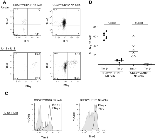
Natural killer (NK) cells are innate lymphocytes that play an important role against viral infections and cancer. This effect is achieved through a complex mosaic of inhibitory and activating receptors expressed by NK cells that ultimately determine the magnitude of the NK-cell response. The T-cell immunoglobulin- and mucin domain-containing (Tim)-3 receptor was initially identified as a T-helper 1-specific type I membrane protein involved in regulating T-cell responses. Human NK cells transcribe the highest amounts of Tim-3 among lymphocytes. Tim-3 protein is expressed on essentially all mature CD56(dim)CD16(+) NK cells and is expressed heterogeneously in the immature CD56(bright)CD16(-) NK-cell subset in blood from healthy adults and in cord blood. Tim-3 expression was induced on CD56(bright)CD16(-) NK cells after stimulation with IL-15 or IL-12 and IL-18 in vitro, suggesting that Tim-3 is a maturation marker on NK cells. Whereas Tim-3 has been used to identify dysfunctional T cells, NK cells expressing high amounts of Tim-3 are fully responsive with respect to cytokine production and cytotoxicity. However, when Tim-3 was cross-linked with antibodies it suppressed NK cell-mediated cytotoxicity. These findings suggest that NK-cell responses may be negatively regulated when NK cells encounter target cells expressing cognate ligands of Tim-3.


Figures

Figure 1
Figure 1
Tim-3 expression on human NK cells. (A) Plots depict Tim-3 expression on NK cells for 3 representative healthy donors. PBMCs were gated on forward angle light scatter (FSC-height and FSC-area) to eliminate doublets, followed by side light scatter (SSC) and FSC gating to define lymphocytes. Dead cells were excluded using an Amine Aqua reactive dye, and CD14 and CD19 staining was used to exclude monocytes and B cells, respectively. CD56 and CD16 were used to identify NK cells within the CD14CD19CD3 population. (B) The plot shows expression of Tim-3 on NK cells in comparison to Tim-3 expression on CD4+ and CD8+ T cells in a representative donor. (C) Graph depicts the frequency (percentage) of Tim-3 on NK cells (solid circles) and CD4+ and CD8+ T cell (open circles) from multiple donors (n = 27). (D) Plot shows NK cells from a healthy donor, gated into CD56brightCD16 and CD56dimCD16+ NK-cell subsets with the expression of Tim-3 on these subsets presented as histograms. Graphs show the frequency (E) and MFI (F) of Tim-3+ CD56brightCD16 (solid circles) and CD56dimCD16+ NK cells (open circles) for 27 healthy individuals. P values were calculated using the Mann-Whitney test. (G) Representative plot shows Tim-3 expression on NK-cell subsets (CD56brightCD16 [gray shade] and CD56dimCD16+ NK cells [black line]) from cord blood obtained at the time of birth. (H) Graph shows Tim-3 expression on NK-cell subsets (CD56brightCD16 [solid circles] and CD56dimCD16+ NK cells [open circles]) from cord blood of 3 healthy newborn infants. (I) Representative plots of Tim-3 coexpression with CD94 on CD56brightCD16 (left) and CD56dimCD16+ (right) NK cells.
Figure 2
Figure 2
Tim-3 expression on NK cells is increased by maturation and cytokine stimulation. (A-B) Plots from a representative donor show the surface expression of Tim-3, on CD56brightCD16 (A) and CD56dimCD16+ (B) NK-cell subsets after culture overnight without stimulation (gray shade) or with IL-2, IL-12 and IL-18, IFN-α, IL-15 and IL-12 (black line for all these stimulations), and IL-15 (dotted line). (C-D) CD19CD14CD3CD56brightCD16 NK cells either lacking or expressing Tim-3 were sorted and incubated for 24 hours with either no stimulation or IL-15. (C) Histograms show Tim-3 expression of sorted cells from a representative donor after culture without (gray shade) or with IL-15 stimulation (black line). (D) Plots depict CD56 and Tim-3 coexpression on sorted CD56brightCD16 Tim-3+ (left) and Tim-3 (right) NK cells without (top) or with (bottom) IL-15 stimulation. These experiments were repeated 3 times with different unrelated healthy donors.
Figure 3
Figure 3
NK cells with the highest surface density of Tim-3 secrete the most IFN-γ after IL-12 and IL-18 stimulation. (A) Plots depict coexpression of Tim-3 and IFN-γ production in unstimulated (top) or stimulated by IL-12 and IL-18 (bottom) NK-cell subsets (CD56brightCD16 [left plots] and CD56dimCD16+ [right plots]) in a representative healthy donor. (B) Graph shows frequency of IFN-γ+ NK cells based on Tim-3 expression on NK-cell subsets (CD56brightCD16 [solid circles] and CD56dimCD16 [open circles]) after IL-12 and IL-18 stimulation in 6 unrelated healthy donors. (C) Histograms show Tim-3 surface expression on IFN-γ (gray shade) and IFNγ+ (black line) cells gated from IL-12– and IL-18–stimulated NK-cell subsets (CD56brightCD16 [left] and CD56dimCD16+ [right]) from a representative donor (n = 6).
Figure 4
Figure 4
NK cells with the highest surface density of Tim-3 degranulate the most when stimulated with target cells. (A) Representative plots depict expression of Tim-3 and surface CD107a in CD56brightCD16 (left) and CD56dimCD16+ (right) NK-cell subsets unstimulated (top) or stimulated (bottom) with the human HLA class I–deficient 721.221 target cells. (B) Graphs shows the frequency of CD107a+ NK cells based on Tim-3 expression on CD56brightCD16 (solid circles) and CD56dimCD16 (open circles) NK-cell subsets, after stimulation with 721.221 cells. Experiment performed with cells from 6 unrelated healthy donors. (C) Histograms depict Tim-3 surface expression on CD107a (gray shade) and CD107a+ (black line) cells gated from IL-12– and IL-18–stimulated NK-cell subsets (CD56brightCD16 [left] and CD56dimCD16+ [right]) from a representative donor (n = 6).
Figure 5
Figure 5
Tim-3 cross-linking inhibits killing by NK cells. (A-C) Each graph depicts the percentage of specific lysis of mouse P815 target cells mediated by the human NK-cell line NKL (A); NKL cells transduced with human Tim-3 containing a FLAG epitope tag on the N terminus, NKL-FLAG-Tim-3 (B); or freshly isolated PBMCs from a representative donor in the absence or presence of the indicated single or combined mAbs: anti–Tim-3 (clone 344801), anti–Tim-3 (clone 344823), anti-NKG2D, anti-CD94, anti-CD16, or anti-FLAG (C). (D-E) Graphs represent lysis of human 721.221 cells transduced with human CD32 Fc receptors by NKL-FLAG-Tim-3 (D) or freshly isolated PBMCs from a representative donor in the presence of the indicated mAbs: anti–Tim-3 (clone 344801), anti–Tim-3 (clone 344823), or anti-CD56 (E). Anti-CD56 was used as a negative control because it does not affect NK-cell cytotoxicity. (F) Graph represents lysis of mouse P815 target cells mediated by PBMCs stimulated overnight with IL-2 from a representative donor in the absence or presence of the indicated single or combined mAbs: anti–Tim-3 (clone 344801), anti–Tim-3 (clone 344823), and anti-CD16. Results are representative of 3 independent experiments performed in triplicate.

Similar articles

Cited by

References

    1. Vilches C, Parham P. KIR: diverse, rapidly evolving receptors of innate and adaptive immunity. Annu Rev Immunol. 2002;20:217–251. - PubMed
    1. Cooper MA, Fehniger TA, Caligiuri MA. The biology of human natural killer-cell subsets. Trends Immunol. 2001;22(11):633–640. - PubMed
    1. Lanier LL, Le AM, Civin CI, Loken MR, Phillips JH. The relationship of CD16 (Leu-11) and Leu-19 (NKH-1) antigen expression on human peripheral blood NK cells and cytotoxic T lymphocytes. J Immunol. 1986;136(12):4480–4486. - PubMed
    1. Fauriat C, Long EO, Ljunggren HG, Bryceson YT. Regulation of human NK-cell cytokine and chemokine production by target cell recognition. Blood. 2010;115(11):2167–2176. - PMC - PubMed
    1. Yu J, Mao HC, Wei M, et al. CD94 surface density identifies a functional intermediary between the CD56bright and CD56dim human NK-cell subsets. Blood. 2010;115(2):274–281. - PMC - PubMed

Publication types

MeSH terms