. 2012 Feb;8(2):e1002540.
doi: 10.1371/journal.ppat.1002540. Epub 2012 Feb 23.

Transient reversal of episome silencing precedes VP16-dependent transcription during reactivation of latent HSV-1 in neurons

Affiliations

Transient reversal of episome silencing precedes VP16-dependent transcription during reactivation of latent HSV-1 in neurons

Ju Youn Kim et al. PLoS Pathog. 2012 Feb.

Abstract

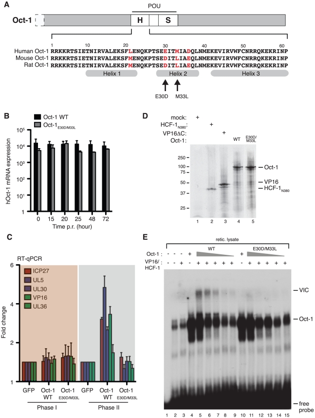
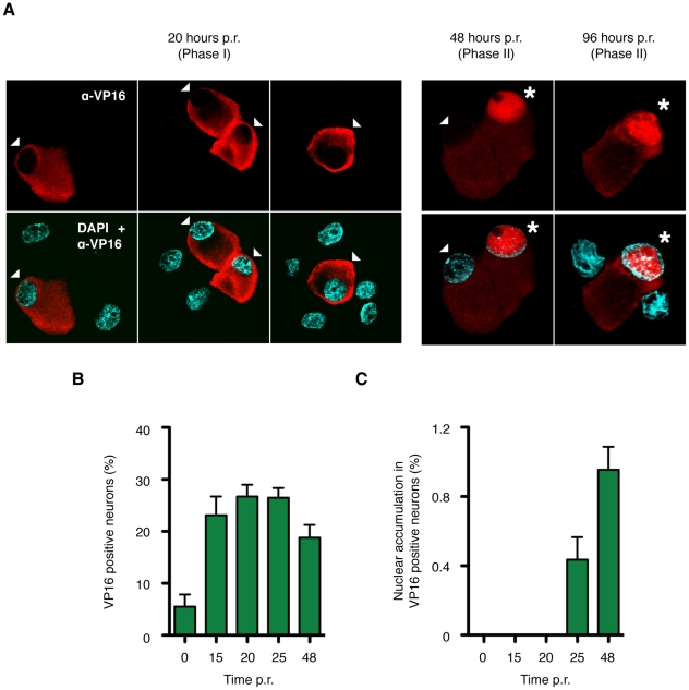
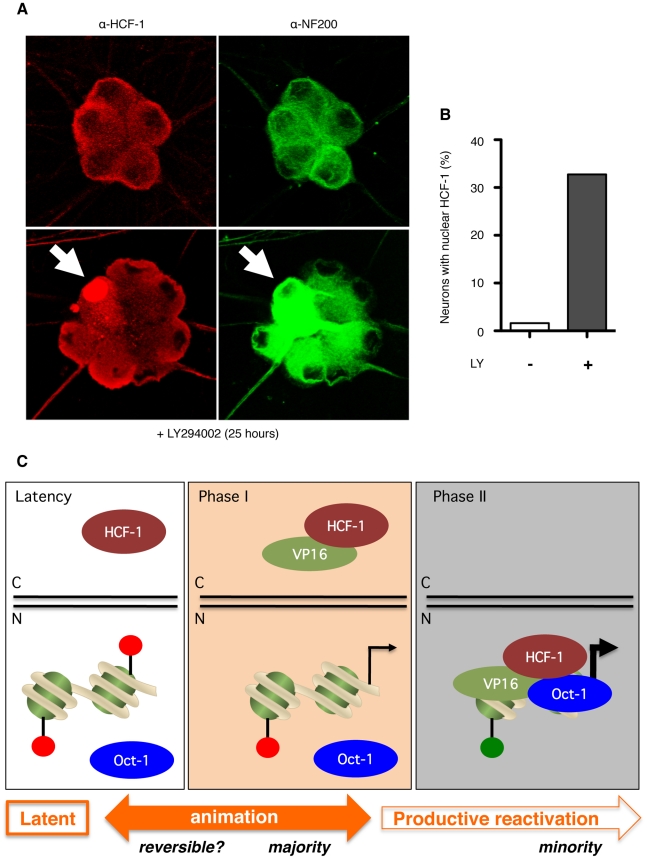
Herpes simplex virus type-1 (HSV-1) establishes latency in peripheral neurons, creating a permanent source of recurrent infections. The latent genome is assembled into chromatin and lytic cycle genes are silenced. Processes that orchestrate reentry into productive replication (reactivation) remain poorly understood. We have used latently infected cultures of primary superior cervical ganglion (SCG) sympathetic neurons to profile viral gene expression following a defined reactivation stimulus. Lytic genes are transcribed in two distinct phases, differing in their reliance on protein synthesis, viral DNA replication and the essential initiator protein VP16. The first phase does not require viral proteins and has the appearance of a transient, widespread de-repression of the previously silent lytic genes. This allows synthesis of viral regulatory proteins including VP16, which accumulate in the cytoplasm of the host neuron. During the second phase, VP16 and its cellular cofactor HCF-1, which is also predominantly cytoplasmic, concentrate in the nucleus where they assemble an activator complex on viral promoters. The transactivation function supplied by VP16 promotes increased viral lytic gene transcription leading to the onset of genome amplification and the production of infectious viral particles. Thus regulated localization of de novo synthesized VP16 is likely to be a critical determinant of HSV-1 reactivation in sympathetic neurons.


Conflict of interest statement

The authors have declared that no competing interests exist.

Figures

Figure 1
Figure 1. Acute replication of HSV-1 in SCG neurons follows the canonical ordered cascade of mRNA accumulation.
(A) Primary neurons were isolated from superior cervical ganglia (SCG) of E21 rats, cultured for 7 days in the presence of 5 µM aphidicolin and 20 µM 5-fluorouracil to eliminate proliferating cells, and then infected with HSV-1 GFP-Us11 at a multiplicity of 3 plaque forming units per neuron (MOI = 3). RNA was collected at 0, 3, 6, 9, and 12 h post-infection (p.i.) and analyzed by quantitative reverse transcription PCR (qRT-PCR) to determine the relative levels of viral immediate-early (ICP27), early (UL30), γ1 leaky-late (VP16) and γ2 true-late (UL36) transcripts. Values represent the average and standard error from the mean from three independent infection experiments. (B) Neuron cultures were treated with the viral DNA polymerase inhibitor phosphonoacetic acid (PAA, 300 µg/ml) for 1 h (hatched bars) or mock treated (filled bars) and then infected with HSV-1 GFP-Us11. Total DNA was prepared at the indicated times and the relative levels of viral genomic DNA determined by quantitative (qPCR) using primers complementary to the HSV-1 UL30 gene. Input DNA was normalized by qPCR detection of the rat RPL19 gene. (C, D) Analysis of γ1 leaky-late (VP16) and γ2 true-late (UL36) transcript levels in the presence or absence of PAA.
Figure 2
Figure 2. During reactivation, HSV-1 exhibits a biphasic profile of viral transcripts in SCG neurons.
(A) Scheme showing a typical reactivation experiment. Neuron cultures were established and then infected with HSV-1 GFP-Us11 (MOI = 1) in the presence of 100 µM acyclovir (ACV). Latency was established over a 7-day period before re-feeding with fresh media lacking ACV. The next day, reactivation was induced with 20 µM LY294002. (B) Profile of viral mRNA accumulation in response to LY294002. RNA was collected at the indicated times and analyzed by qRT-PCR. Values are normalized against the 0 h sample [ICP27, 171 copies/sample; UL5, 135 copies/sample; UL30, 94 copies/sample; VP16, 347 copies/sample and UL36 130 copies/sample]. Data is derived from three or more independent cultures and reactivation experiments. (C) Reactivation profiling in the presence of the viral DNA encapsidation inhibitor WAY150138 (20 µg/ml). (D) Transcript levels at 20 h post induction in the absence (−) or presence (+) of protein synthesis inhibitor cyclohexamide (CHX, 10 µg/ml). To ensure cell viability, CHX was added 10 h after LY294002, prior to the appearance of new viral transcripts.
Figure 3
Figure 3. HSV-1 GFP-Us11 replication occurs in Phase II of reactivation.
Latent cultures of SCG cells were induced with LY294002 as described before. (A) Viral DNA Content was determined by qPCR at different times post-reactivation (p.r.) in the absence or presence of 300 µg/ml PAA. (B) Quantitative RT-PCR analysis of VP16 and UL36 mRNA levels in the absence or presence of 300 µg/ml PAA during Phase I (15–20 h) and in Phase II (48 h). (C) Number of infectious virus particles was determined by plaque assay.
Figure 4
Figure 4. Transactivation function of VP16 is required during Phase II.
(A) Structure of VP16 showing the 12-bp insertion (in1814) between the structured N-terminal domain and the C-terminal activation domain (AD) that disrupts VP16-induced complex assembly . (B) SCG neurons were infected with mutant (in1814) or marker rescue (in1814R) viruses (MOI = 1) in the presence of 100 µM acyclovir and maintained for 7 days before measuring the relative amounts of viral genomic DNA by qPCR. (C) Reactivation was induced with 20 µM LY294002 in media lacking ACV and maintained for 7 days before harvest and plaque assay to detect infectious virus. (D) Comparison of viral transcript levels during reactivation by in1814 (‘M’) and in1814R (‘R’) at 15 and 20 h post-induction (Phase I) and at 72 h post-induction (Phase II). For each time point, transcript levels from the in1814 (‘M’) sample were set to 1 and the value for the corresponding transcript from in1814R (‘R’) plotted as the fold difference. (E) Depletion of VP16 using RNA interference. Latently infected neuron cultures were infected with a lentivirus expressing a VP16-specific short-hairpin RNA [shRNA] (KD) or with a control lentivirus (Con). ShRNAs were allowed to accumulate for 5 days before reactivation was induced with LY294002 and allowed to proceed for 5 days in media lacking ACV. Lysates were prepared and probed by immunoblotting to detect VP16 and the loading control, Rho-GDI. (F) Quantitation of infectious virus by plaque assay. (G) Comparison of viral transcript levels in the absence of VP16. Values from the control culture are plotted relative to the corresponding value from the VP16 shRNA (KD) culture.
Figure 5
Figure 5. Elevated Phase II viral transcript levels in neurons expressing human Oct-1.
(A) Schematic of Oct-1 showing the location of the POU DNA-binding domain near the middle of the protein and an alignment of the human, mouse and rat POU homeo (POUH) subdomain sequences. The four variable positions are located in helix-1 and helix-2 and are numbered according to their position within the POUH sequence. A rodent-like Oct-1 derivative (Oct-1E30D/M33L) was constructed by changing glutamic acid-30 and methionine-33 to the aspartic acid and leucine of the mouse/rat sequence. (B) SCG neuron cultures were infected with HSV-1 GFP-Us11, maintained for 5 days in the presence of ACV and then infected with lentiviral vectors encoding GFP or human Oct-1. After a further 5 days, ACV was removed and reactivation induced with 20 µM LY294002. RNA was collected at intervals and analyzed by qRT-PCR using human Oct-1 specific primers. For each time point, values for each transcript were compared to those from the GFP-expressing neurons (set to 1.0). (C) Relative levels of viral transcripts (ICP27, UL5, UL30, VP16 and UL36) after reactivation of HSV-1 GFP-Us11 in neurons expressing GFP, wild type human Oct-1 (WT) and human Oct-1E30D/M33L (MUT). (D) Wild type (WT) and E30D/M33L (MUT) versions of human Oct-1, VP16 (residues 5–412, VP16ΔC) and the β-propeller domain of human HCF-1 (residues 1–380, HCF-1N380) were synthesized by in vitro translation in the presence of 35S-methionine and visualized by 10% SDS-PAGE followed by autoradiography. (E) Assembly of the VP16-induced complex (VIC) by rodent-like (E30D/M33L) Oct-1 is greatly reduced compared to wild type human Oct-1. Recombinant Oct-1, VP16 and HCF-1 proteins were assayed for VIC formation by gel shift assay using a 32P-labeled probe containing an (OCTA+)TAATGARAT element from the HSV-1 ICP0 promoter . The first three lanes are controls showing probe alone (lane 1), un-programmed rabbit reticulocyte lysate (lane 2), and a mix of lysates containing recombinant VP16 and HCF-1 (lane 3). The shift formed by the rabbit Oct-1 present in the lysate is greatly enhanced by the presence of either wild type or mutant human Oct-1 (lanes 4 and 10). A slower migrating complex (VIC) is formed by addition of VP16 and HCF-1 in the presence of wild type Oct-1 (lane 5) and only weakly by the mutant (lane 11). Reducing the amount of wild type Oct-1 by 5, 10, 50 and 100-fold respectively (lanes 6–9) reduces but does not eliminate this complex. No VIC is detected over a similar range of using mutant Oct-1.
Figure 6
Figure 6. VP16 is expressed during Phase I but is localized to the cytoplasm.
(A) Detection of VP16 in latently infected neurons by indirect immunofluorescence microscopy using an α-VP16 rabbit polyclonal antibody (red). Individual nuclei were visualized with DAPI (blue). Three representative fields are shown from cover slips collected at 20 h post-induction (Phase I) and single fields from 48 and 96 h (Phase II). An arrowhead is used to indicate the cell body of neurons with a predominantly cytoplasmic VP16 signal, whereas those displaying a strong nuclear VP16 signal are indicated with an asterisk. Neurons that do not express VP16 are left unmarked but are evident from the DAPI stained nuclei. (B) Quantitation of the immunofluorescence analysis. Numbers of VP16 positive neurons are expressed as a percentage of all neurons scored. Data is compiled from three independent reactivation experiments. Between 500 and 3,000 neurons were scored per cover slip. (C) Percentage of VP16 positive neurons with a predominantly nuclear rather than cytoplasmic α-VP16 signal.
Figure 7
Figure 7. Relocalization of HCF-1 in response to PI3-kinase inhibition.
(A) Confocal image showing a cluster of neuronal cell bodies probed with α-HCF-1 rabbit polyclonal antibody (red) and α-neurofilament-200 (NF200) mouse monoclonal antibody (green) at 25 h post treatment with LY294002 (lower panels) or vehicle (upper panels). Note HCF-1 signal throughout the cell body and axonal processes with sparing in the nucleus. Nuclear accumulation is detected in one neuron (arrow) in the LY294002-treated sample. (B) Quantitation of neurons showing obvious nuclear HCF-1 at 25 h post treatment with LY294002 (+) or vehicle (−) (C) Model for the two-step reactivation program observed in sympathetic neurons induced by inhibition of NGF signaling. The latent HSV-1 episome resides in the nucleus (‘N’) and is associated with histones carrying post-translational modifications typical of repressed chromatin (red lollipops). HCF-1 is sequestered in the cytoplasm, whereas Oct-1 is presumed to be nuclear throughout. Host-mediated alterations to episome chromatin during Phase I allows for generalized transcription of viral lytic genes. VP16 is synthesized, but accumulates in the cytoplasm (‘C’), possibly through association with HCF-1. We speculate that a smaller number of animation events advance to Phase II, coincident with the accumulation of VP16 and HCF-1 into the nucleus permitting the assembly of the VP16-induced complex on viral IE promoters in association with Oct-1. This leads to the recruitment of additional chromatin modifiers that apply chromatin marks associated with active transcription (green lollipops).

Similar articles

Cited by

References

    1. Roizman B. The checkpoints of viral gene expression in productive and latent infection: The role of the HDAC/CoREST/LSD1/REST repressor complex. J Virol. 2011;855:7474–7482. - PMC - PubMed
    1. Penkert RR, Kalejta RF. Tegument protein control of latent herpesvirus establishment and animation. Herpesviridae. 2011;2:3. - PMC - PubMed
    1. Knipe DM, Cliffe A. Chromatin control of herpes simplex virus lytic and latent infection. Nat Rev Microbiol. 2008;6:211–221. - PubMed
    1. Bloom DC, Giordani NV, Kwiatkowski DL. Epigenetic regulation of latent HSV-1 gene expression. Biochim Biophys Acta. 2010;1799:246–256. - PMC - PubMed
    1. Stevens JG, Wagner EK, Devi-Rao GB, Cook ML, Feldman LT. RNA complementary to a herpesvirus alpha gene mRNA is prominent in latently infected neurons. Science. 1987;235:1056–1059. - PubMed

Publication types

MeSH terms

Substances