. 2012 Feb;8(2):e1002543.
doi: 10.1371/journal.ppat.1002543. Epub 2012 Feb 23.

The invasive capacity of HPV transformed cells requires the hDlg-dependent enhancement of SGEF/RhoG activity

Affiliations

The invasive capacity of HPV transformed cells requires the hDlg-dependent enhancement of SGEF/RhoG activity

Vanitha Krishna Subbaiah et al. PLoS Pathog. 2012 Feb.

Abstract

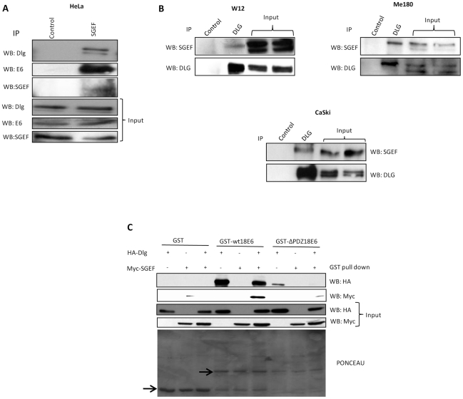
A major target of the HPV E6 oncoprotein is the human Discs Large (hDlg) tumour suppressor, although how this interaction contributes to HPV-induced malignancy is still unclear. Using a proteomic approach we show that a strong interacting partner of hDlg is the RhoG-specific guanine nucleotide exchange factor SGEF. The interaction between hDlg1 and SGEF involves both PDZ and SH3 domain recognition, and directly contributes to the regulation of SGEF's cellular localization and activity. Consistent with this, hDlg is a strong enhancer of RhoG activity, which occurs in an SGEF-dependent manner. We also show that HPV-18 E6 can interact indirectly with SGEF in a manner that is dependent upon the presence of hDlg and PDZ binding capacity. In HPV transformed cells, E6 maintains a high level of RhoG activity, and this is dependent upon the presence of hDlg and SGEF, which are found in complex with E6. Furthermore, we show that E6, hDlg and SGEF each directly contributes to the invasive capacity of HPV-16 and HPV-18 transformed tumour cells. These studies demonstrate that hDlg has a distinct oncogenic function in the context of HPV induced malignancy, one of the outcomes of which is increased RhoG activity and increased invasive capacity.


Conflict of interest statement

The authors have declared that no competing interests exist.

Figures

Figure 1
Figure 1. Identification of SGEF as an interacting partner of Dlg.
Panel A. HEK293 cells were transfected with HA-tagged Dlg expression plasmid and after 24 hrs cells were immunoprecipitated using anti-HA antibody-conjugated agarose beads. The total protein complex was then subjected to mass spectroscopy analysis. The table shows a selection of the prominent Dlg interacting proteins, together with the peptide sequences showing SGEF as a novel interacting partner. Panel B. HEK293 cells were transfected with HA-tagged Dlg and Flag-tagged SGEF expression plasmids, as indicated, and after 24 hrs cell extracts were immunoprecipitated using anti HA-antibody conjugated agarose beads. Dlg-bound SGEF was then detected by western blotting using anti-Flag antibody. Panel C. Mouse liver lysate was incubated with streptavidin-Sepharose beads coupled with biotinylated peptides corresponding to the last 10 amino acids of SGEF or control (CTRL) peptides (i) in which the last 4 amino acids were replaced by glycine (underlined). (ii) After being washed, the samples were separated by SDS-PAGE and immunobloted with anti-Dlg1 antibodies. Panel D. HaCaT and HEK293 cell extracts were subjected to immunoprecipitation with pre-immune, anti-SGEF or anti-Dlg antibody as indicated, and the corresponding co-immunoprecipitating Dlg or SGEF detected by western blotting as indicated. As an additional control, HEK293 cells were also transfected with siRNA SGEF or siRNA Luc 72 hrs prior to harvesting, thereby confirming the identity of the SGEF protein detected with the anti SGEF antibody.
Figure 2
Figure 2. The interaction between Dlg and SGEF involves PBM-PDZ and SH3-SH3 domain recognition.
Panel A. Diagrammatic representation of the Dlg deletion mutants used in this study. Panel B. HEK 293 cells were transfected with Flag-tagged SGEF expression plasmid and cell extracts made after 24 hrs. These were then incubated with a panel of GST.Dlg fusion proteins encompassing different domains of the Dlg protein and GST alone as a negative control. Bound SGEF was then detected by western blotting using anti-Flag antibody (upper panel). The middle panel shows the Ponceau stain of the membrane confirming similar levels of GST protein expression, whilst the bottom panel shows the input of SGEF used in each assay. Panel C. HEK293 cells were transfected with HA-tagged wild type Dlg and two mutants encompassing the N and C terminal halves of the protein. After 24 hrs the cells were extracted and Dlg precipitated using anti-HA antibody. The co-immunoprecipitated endogenously expressed SGEF was then detected by western blot using anti-SGEF antibody (upper left panel). Input proteins are shown in the two right panels and the lower left panel shows the immunoprecipitated Dlg (indicated by arrows). Panel D. HEK293 cells were transfected with myc-tagged wild type SGEF and mutants deleted in the PDZ binding motif and the SH3 domain. After 24 hrs cell extracts were made and incubated with purified GST fusion protein, consisting of GST alone, wild type Dlg and the N and C terminal halves of Dlg. Bound SGEF was detected by western blot using anti-myc antibody (upper panel). The lower panel shows the Ponceau stain of the membrane demonstrating the levels of GST protein expression with arrows indicating the relevant full-length GST fusion proteins. Panel E. Schematic summarizing the results of the interaction assays, demonstrating interaction between the Dlg PDZ1 and PDZ2 domains with the SGEF PBM plus association between the SGEF and Dlg SH3 domains.
Figure 3
Figure 3. Dlg regulates the cellular localisation of SGEF.
Panel A. HEK293 cells were transfected with myc-tagged SGEF expression plasmid together with increasing amounts of HA-tagged Dlg expression plasmid. After 24 hrs cells were extracted into soluble (left panel) and insoluble (right panel) fractions. Changes in the patterns of SGEF localisation were detected by western blot using anti-Myc antibody and Dlg was detected using anti-HA antibody. The levels of β-gal expression are also shown as a marker for transfection efficiency and loading control. Panel B. HEK293 cells were transfected with the Myc-tagged SGEF expression plasmids together with a HA-tagged Dlg expression plasmid. After 24 hrs the cells were extracted into soluble and insoluble fractions and the pattern of expression determined by western blot analysis. β-gal expression was used as a marker for transfection efficiency and a loading control. Panel C. HaCaT cells stably selected with control targeting plasmid (TR2), shDlg targeting plasmid, and cells targeted for shDlg but rescued with rat Dlg were analysed for the pattern of SGEF expression in the soluble and insoluble fractions by western blotting with anti-SGEF antibody. α-Actinin was used as a loading control. Note the complete loss of expression of SGEF from the insoluble fraction of the cell upon hDlg ablation. Panel D. Western blot analysis of SGEF expression levels in the total cell lysates prepared from stable cell lines used in Panel C.
Figure 4
Figure 4. Dlg recruits SGEF to the cytoskeletal network.
Panel A. HEK293 cells were transfected with HA-tagged Dlg and Flag-tagged SGEF expression plasmids as indicated and after 24 hrs cells were extracted and divided into cytosolic, membrane, nuclear and cytoskeletal fractions. The pattern of Dlg and SGEF expression was then determined by western blotting with anti-HA and anti-Flag antibodies respectively. Loading controls confirming the integrity of the differential extractions are α-tubulin, E-cadherin, p84 and vimentin for the cytosolic, membrane, nuclear and cytoskeletal fractions respectively. Panel B. HaCaT cells transfected with HA-Dlg, Flag-SGEF or co-transfected with the two cDNAs were fixed and processed for immunofluorescence with anti-Flag to detect SGEF and anti-HA to detect Dlg. i) Single HA-Dlg transfection. ii) Single Flag-SGEF transfection. iii) Co-transfection with Dlg in red and SGEF in green. The arrows indicate discrete areas of co-localisation within cytoplasmic and membrane sites.
Figure 5
Figure 5. Dlg enhances RhoG activity in an SGEF dependent manner.
Panel A. HEK293 cells were either transfected with vector or HA-tagged Dlg and Flag-tagged SGEF as indicated. After 24 hrs cell extracts were made which were then incubated with purified GST-ELMO to pull down active RhoG which was detected by western blot analysis. The three lower panels show total protein inputs for RhoG, Dlg and SGEF. Panel B. Graph showing the quantification from multiple GST-ELMO pull-downs, showing the fold change in the levels of RhoG activity under the different experimental conditions. Error bars represent ±SD of three independent experiments Panel C. Extracts from HaCaT cells stably ablated for hDlg expression (shDlg) either with or without Dlg rescue expression were used in a GST-ELMO pulldown assay to determine RhoG activity. Extracts from untreated HaCaT cells or HaCaT cells stably expressing control shRNA(TR2) were used as control.
Figure 6
Figure 6. High levels of RhoG activity in HPV-18 transformed cells is hDlg and HPV -dependent.
Panel A. HeLa cells were transfected with siRNAs against luciferase (Luc) control, E6/E7, hDlg or SGEF as indicated. After 72 hrs cell extracts were then incubated with purified GST.ELMO to determine the levels of active RhoG, which was detected by western blotting (upper panel). The lower panels show input levels of total RhoG, hDlg, SGEF, p53 and α-Actinin. Also shown is the Ponceau stain of the membrane showing constant levels of GST.ELMO. The graph shows the quantifications from multiple GST.ELMO pull-downs, and shows the fold change in the levels of RhoG activity under the different experimental conditions. Error bars represent +/− SD of four independent experiments. Note the modest decrease in active RhoG following the removal of E6/E7 and the dramatic decrease following removal of hDlg. Panel B. HeLa cells were transfected with control siRNA (Luc) siRNA to E6/E7 (i) or siRNA to hDlg-1 (ii) and were then analysed for the levels of hDlg, SGEF, and p53 expression after 72 hrs in the insoluble and soluble compartments of the cell. α-Actinin was used as a loading control. Note the marked increase in hDlg levels in the soluble fraction with concomitant decrease in the insoluble fraction following E6/E7 removal, which is also accompanied by a decrease in SGEF levels in this compartment and a similar loss of SGEF in the insoluble compartment is seen following siRNA ablation of hDlg-1 expression. Panel C. HeLa cells were transfected with control siRNA (Luc), siRNA to E6 (si18E6intronic) or siRNA to E6/E7 and were then analysed for levels of expression of SGEF and p53 in total cell lysates after 72 hrs. α-Actinin was used as a loading control.
Figure 7
Figure 7. HPV-18E6, hDlg and SGEF exist as a complex.
Panel A. HeLa cells were seeded in 10 cm2 dishes. After 24 hrs, cell extracts were prepared and immunoprecipitated using either the control antibody or the SGEF antibody. hDlg and HPV18E6 bound to the SGEF were detected using the anti-Dlg and anti-E6 antibodies respectively. The immunoprecipitated SGEF was detected using anti-SGEF antibody. The bottom 3 lanes show the input levels for hDlg, HPV18E6 and SGEF used in this assay. Panel B. HPV positive W12, Me180 and CaSki cells were seeded on 10 cm2 dishes and cellular extracts immunoprecipitated using either control antibody or anti-hDlg-1 antibody. The Dlg bound SGEF was detected by western blotting with the anti-SGEF antibody and the immunoprecipitated Dlg was detected using the anti-Dlg antibody. The right two lanes in each panel show the input levels (20%) of SGEF and Dlg used in the control and Dlg immunoprecipitates. Panel C. HEK 293 cells were transfected with HA-tagged Dlg and Myc-tagged SGEF expression plasmids either alone or in combination and the cell extracts made after 24 hrs. These were then incubated with GST-wt18E6 or GST-ΔPDZ18E6 and GST alone as a negative control. Bound Dlg was then detected by western blotting using anti-HA antibody (upper panel), bound SGEF was detected using anti-Myc antibody (second panel). The third and fourth panels shows the input of Dlg and SGEF used in this assay. The bottom panel shows the Ponceau stain of the membrane showing the levels of GST protein expression.
Figure 8
Figure 8. HPV18E6 does not degrade the NP-40 insoluble pool of hDlg in the presence of SGEF.
HEK293 cells were transfected with HA-tagged Dlg, Myc-tagged SGEF, untagged HPV18E6 expression plasmids either alone or in different combinations as indicated. After 24 hrs the cells were extracted into NP-40 soluble and NP-40 insoluble fractions and the patterns of expressions of Dlg, SGEF and E6 determined by western blot analysis. β-gal expression was used as a marker for transfection efficiency and a loading control.
Figure 9
Figure 9. The hDlg/SGEF module is required for the invasive capacity of HeLa and CaSki cells.
The cells (HeLa in Panel A and CaSki in Panel B) were transfected with siRNAs to Luciferase (Luc), E6/E7, hDlg or SGEF and after 72 hrs the cells were harvested and equal numbers plated onto Matrigel invasion chambers. After overnight incubation the numbers of invading cells in the lower chamber were counted. The graphs show the fold change in the numbers of invading cells from multiple assays, where siLuc- transfected cells were scored as the reference point. Error bars represent ±SD of multiple experiments. The right hand panels show the western blot analysis of the levels of expression in total cell extracts of hDlg, p53 and SGEF following siRNA transfections performed in parallel with the invasion assays. α-Actinin is shown as the loading control.

Similar articles

Cited by

References

    1. zur Hausen H. Immortalization of human cells and their malignant conversion by high-risk human papillomavirus genotypes. Semin Cancer Biol. 1999;9:405–411. - PubMed
    1. zur Hausen H. Papillomaviruses and cancer: from basic studies to clinical application. Nat Rev Cancer. 2002;2:342–50. - PubMed
    1. Barbosa MS, Schlegel R. The E6 and E7 genes of HPV-18 are sufficient for inducing two-stage in vitro transformation of human keratinocytes. Oncogene. 1989;4:1529–1532. - PubMed
    1. Hawley-Nelson P, Vousden KH, Hubbert NL, Lowy DR, Schiller JT. HPV-16 E6 and E7 proteins cooperate to immortalise human foreskin keratinocytes. EMBO J. 1989;8:3905–3910. - PMC - PubMed
    1. Song S, Liem A, Miller JA, Lambert PF. Human papillomavirus type 16 E6 and E7 contribute differently to carcinogenesis. Virology. 2000;267:141–150. - PubMed

Publication types

MeSH terms

Substances

LinkOut - more resources