The ebola virus interferon antagonist VP24 directly binds STAT1 and has a novel, pyramidal fold
- PMID: 22383882
- PMCID: PMC3285596
- DOI: 10.1371/journal.ppat.1002550
The ebola virus interferon antagonist VP24 directly binds STAT1 and has a novel, pyramidal fold
Erratum in
- PLoS Pathog. 2013 Dec;9(12). doi:10.1371/annotation/360ddc68-0313-4eae-af24-043cc040c52d
Abstract
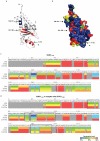
Ebolaviruses cause hemorrhagic fever with up to 90% lethality and in fatal cases, are characterized by early suppression of the host innate immune system. One of the proteins likely responsible for this effect is VP24. VP24 is known to antagonize interferon signaling by binding host karyopherin α proteins, thereby preventing them from transporting the tyrosine-phosphorylated transcription factor STAT1 to the nucleus. Here, we report that VP24 binds STAT1 directly, suggesting that VP24 can suppress at least two distinct branches of the interferon pathway. Here, we also report the first crystal structures of VP24, derived from different species of ebolavirus that are pathogenic (Sudan) and nonpathogenic to humans (Reston). These structures reveal that VP24 has a novel, pyramidal fold. A site on a particular face of the pyramid exhibits reduced solvent exchange when in complex with STAT1. This site is above two highly conserved pockets in VP24 that contain key residues previously implicated in virulence. These crystal structures and accompanying biochemical analysis map differences between pathogenic and nonpathogenic viruses, offer templates for drug design, and provide the three-dimensional framework necessary for biological dissection of the many functions of VP24 in the virus life cycle.
Conflict of interest statement
The authors have declared that no competing interests exist.
Figures


Similar articles
-
The ebolavirus VP24 interferon antagonist: know your enemy.Virulence. 2012 Aug 15;3(5):440-5. doi: 10.4161/viru.21302. Epub 2012 Aug 15. Virulence. 2012. PMID: 23076242 Free PMC article.
-
VP24-Karyopherin Alpha Binding Affinities Differ between Ebolavirus Species, Influencing Interferon Inhibition and VP24 Stability.J Virol. 2017 Jan 31;91(4):e01715-16. doi: 10.1128/JVI.01715-16. Print 2017 Feb 15. J Virol. 2017. PMID: 27974555 Free PMC article.
-
Ebola virus VP24 proteins inhibit the interaction of NPI-1 subfamily karyopherin alpha proteins with activated STAT1.J Virol. 2007 Dec;81(24):13469-77. doi: 10.1128/JVI.01097-07. Epub 2007 Oct 10. J Virol. 2007. PMID: 17928350 Free PMC article.
-
Ebolavirus interferon antagonists-protein interaction perspectives to combat pathogenesis.Brief Funct Genomics. 2018 Nov 26;17(6):392-401. doi: 10.1093/bfgp/elx034. Brief Funct Genomics. 2018. PMID: 29140409 Review.
-
Insights into Ebola Virus VP35 and VP24 Interferon Inhibitory Functions and their Initial Exploitation as Drug Targets.Infect Disord Drug Targets. 2019;19(4):362-374. doi: 10.2174/1871526519666181123145540. Infect Disord Drug Targets. 2019. PMID: 30468131 Review.
Cited by
-
Intracellular Ebola virus nucleocapsid assembly revealed by in situ cryo-electron tomography.Cell. 2024 Oct 3;187(20):5587-5603.e19. doi: 10.1016/j.cell.2024.08.044. Epub 2024 Sep 17. Cell. 2024. PMID: 39293445
-
New Perspectives on the Biogenesis of Viral Inclusion Bodies in Negative-Sense RNA Virus Infections.Cells. 2021 Jun 10;10(6):1460. doi: 10.3390/cells10061460. Cells. 2021. PMID: 34200781 Free PMC article. Review.
-
Jamaican fruit bats' competence for Ebola but not Marburg virus is driven by intrinsic differences.Nat Commun. 2025 Mar 25;16(1):2884. doi: 10.1038/s41467-025-58305-4. Nat Commun. 2025. PMID: 40133326 Free PMC article.
-
Global phosphoproteomic analysis of Ebola virions reveals a novel role for VP35 phosphorylation-dependent regulation of genome transcription.Cell Mol Life Sci. 2020 Jul;77(13):2579-2603. doi: 10.1007/s00018-019-03303-1. Epub 2019 Sep 28. Cell Mol Life Sci. 2020. PMID: 31562565 Free PMC article.
-
The Role of Ebola Virus VP24 Nuclear Trafficking Signals in Infectious Particle Production.bioRxiv [Preprint]. 2024 Mar 13:2024.03.13.584761. doi: 10.1101/2024.03.13.584761. bioRxiv. 2024. PMID: 38559040 Free PMC article. Preprint.
References
-
- Retuya TJA, Miranda MEG, Ksiazek TG, Khan AS, Sanchez A, et al. Is the ebola-reston virus a threat to occupationally exposed humans? J Clin Epidemiol. 1997;50:32S.
-
- Sanchez A, Khan AS, Zaki SR, Nabel GJ, Ksiazek TG, et al. Filoviridae: Marburg and Ebola Viruses. In: Fields BN, Knipe DM, Howley PM, Griffin DE, Lamb RA, et al., editors. Fields virology. 4 ed. Philadelphia: Lippincott Williams & Wilkins; 2001. pp. 1279–1304.
-
- Baize S, Leroy EM, Georges-Courbot MC, Capron M, Lansoud-Soukate J, et al. Defective humoral responses and extensive intravascular apoptosis are associated with fatal outcome in Ebola virus-infected patients. Nat Med. 1999;5:423–426. - PubMed
Publication types
MeSH terms
Substances
Grants and funding
- NS070899/NS/NINDS NIH HHS/United States
- AI081982/AI/NIAID NIH HHS/United States
- R01 AI081982/AI/NIAID NIH HHS/United States
- R01 GM020501/GM/NIGMS NIH HHS/United States
- S10 RR029388/RR/NCRR NIH HHS/United States
- R43 AI1088843/AI/NIAID NIH HHS/United States
- 5T32 NS041219/NS/NINDS NIH HHS/United States
- GM020501/GM/NIGMS NIH HHS/United States
- T32 AI007244/AI/NIAID NIH HHS/United States
- R01 NS070899/NS/NINDS NIH HHS/United States
- GM093325/GM/NIGMS NIH HHS/United States
- R01 GM093325/GM/NIGMS NIH HHS/United States
- F32 GM020501/GM/NIGMS NIH HHS/United States
- GM066170/GM/NIGMS NIH HHS/United States
- AI2008031/AI/NIAID NIH HHS/United States
- RR029388/RR/NCRR NIH HHS/United States
- T32 NS041219/NS/NINDS NIH HHS/United States
- R01 GM066170/GM/NIGMS NIH HHS/United States
LinkOut - more resources
Full Text Sources
Medical
Molecular Biology Databases
Research Materials
Miscellaneous

