. 2012;8(2):e1002481.
doi: 10.1371/journal.pgen.1002481. Epub 2012 Feb 23.

A pathogenic mechanism in Huntington's disease involves small CAG-repeated RNAs with neurotoxic activity

Affiliations

A pathogenic mechanism in Huntington's disease involves small CAG-repeated RNAs with neurotoxic activity

Mónica Bañez-Coronel et al. PLoS Genet. 2012.

Abstract

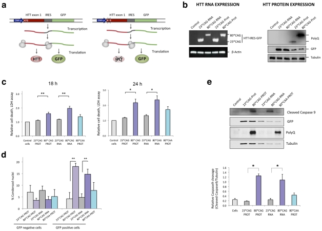
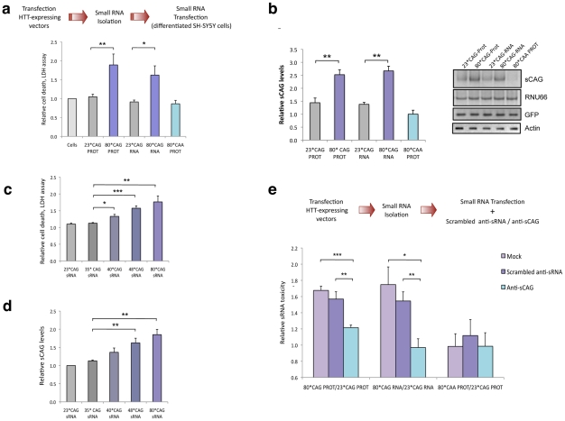
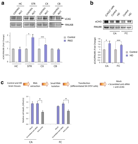
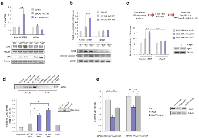
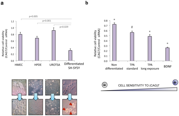
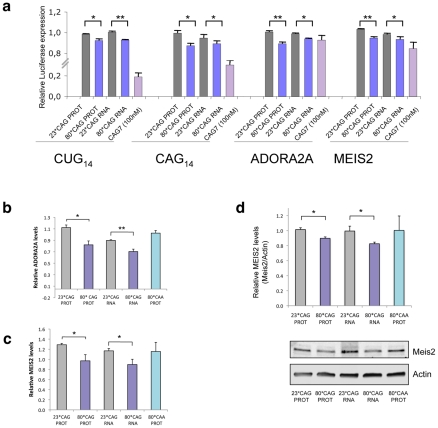
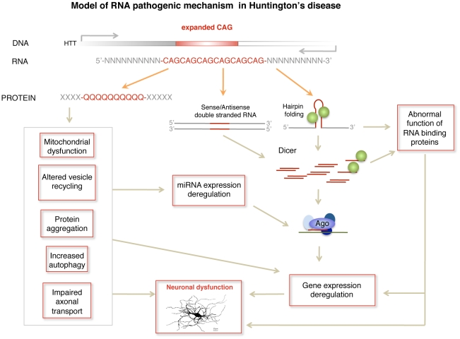
Huntington's disease (HD) is an autosomal dominantly inherited disorder caused by the expansion of CAG repeats in the Huntingtin (HTT) gene. The abnormally extended polyglutamine in the HTT protein encoded by the CAG repeats has toxic effects. Here, we provide evidence to support that the mutant HTT CAG repeats interfere with cell viability at the RNA level. In human neuronal cells, expanded HTT exon-1 mRNA with CAG repeat lengths above the threshold for complete penetrance (40 or greater) induced cell death and increased levels of small CAG-repeated RNAs (sCAGs), of ≈21 nucleotides in a Dicer-dependent manner. The severity of the toxic effect of HTT mRNA and sCAG generation correlated with CAG expansion length. Small RNAs obtained from cells expressing mutant HTT and from HD human brains significantly decreased neuronal viability, in an Ago2-dependent mechanism. In both cases, the use of anti-miRs specific for sCAGs efficiently blocked the toxic effect, supporting a key role of sCAGs in HTT-mediated toxicity. Luciferase-reporter assays showed that expanded HTT silences the expression of CTG-containing genes that are down-regulated in HD. These results suggest a possible link between HD and sCAG expression with an aberrant activation of the siRNA/miRNA gene silencing machinery, which may trigger a detrimental response. The identification of the specific cellular processes affected by sCAGs may provide insights into the pathogenic mechanisms underlying HD, offering opportunities to develop new therapeutic approaches.


Conflict of interest statement

The authors have declared that no competing interests exist.

Figures

Figure 1
Figure 1. CAG-expanded exon 1 of human HTT is toxic at the RNA level.
A. CAG-unexpanded (wild-type; 23 CAG repeats) and CAG-expanded (mutant; 80 CAG repeats) constructs of human HTT exon 1 (HTT exon 1) were subcloned into a pIRES-EGFP vector. Each variant was produced as a normal translated form (left) and a form lacking the translation initiation codon (right). The specific role of the expanded protein was analyzed with a construct expressing CAA-expanded HTT-e1. The use of IRES-based bicistronic vectors with a GFP reporter allows monitoring of transfected cells. B. The four different constructs express the mRNA HTT-IRES-GFP (left) and the GFP reporter protein (right). HTT protein is only expressed in the constructs containing the ATG translation initiation codon (right). C. Differentiated SH-SY5Y cells were transfected with the HTT-IRES-GFP vectors and LDH cell toxicity assay was performed 18 h and 24 h after transfection. Expression of CAG-expanded HTT (RNA or protein) resulted in dramatic cell death. CAA-expanded HTT-e1 didn't induce a significant effect on cell viability at the time points analyzed (n = 4; *p<0.05, **p<0.01, ***p<0.001). D. The percentage of dead transfected cells was also determined 36 hours after transfection by counting 200 GFP-negative cells (left) and 200 GFP-positive cells (right), scoring in each case the presence of nuclear fragmentation. Values represent the percentage of cells showing nuclear condensation in each situation ± SD (n = 3; **p<0.01). E. Expression of CAG-expanded HTT RNA induced caspase 9 cleavage. GFP blots highlight the expression of all constructs in transfected cells and polyglutamine (PolyQ) blots show expression of expanded HTT protein. Densitometry determinations of cleaved caspase 9 vs. α-Tubulin were performed on cells lysated 24 hours after transfection. Results are presented as the mean of arbitrary optical density units (O.D. units ± SEM; n = 3; *p<0.05, ***p<0.001). In C. and E., values represent the mean fold change with respect to the control non-transfected cells ± SEM.
Figure 2
Figure 2. Expanded HTT generates CAG-repeated sRNAs with toxic activity.
A. sRNA fraction (<100 nt) were isolated from cells expressing HTT-e1 constructs and equal amounts of each pool were transfected. Both 80*CAG-RNA and 80*CAG-PROT- derived sRNA pools induced death of differentiated SH-SY5Y cells (n = 5; *p<0.05,**p<0.01). 80*CAA-PROT-derived sRNA pools didn't affect SH-SY5Y cell viability. B. The expression of CAG-expanded HTT leads to an increase in CAG-repeated sRNAs of ∼21-nt (sCAG). sCAG levels were quantified using RNU66 as the reference sRNA, and normalized with respect to GFP expression, which indicates the percentage on transfected living cells 24 hours after transfection (n = 4; **p<0.01). C. HTT sRNA toxicity correlates with the length of the CAG expansion, distinguishing pathogenic and non-pathogenic number of CAG repeats (n = 4; * p<0.05, **p<0.01 ***p<0.001. D. HTT sRNA toxicity correlates with the generation of sCAG species (n = 4; **p<0.01). E. Anti-(CAG)7 sRNA (anti-sCAG) prevents cell damage caused by mutant-HTT-derived sRNA pool. Control sRNA inhibitors did not mitigate sRNA HTT toxicity (n = 4; *p<0.05, **p<0.01, **p<0.001, determinations were performed in quintuplicates). Values represent mean of the ratio expanded-HTT sRNA toxicity vs non-expanded-HTT sRNA toxicity ± SEM. In A. B. C. and D. values represent the mean fold change with respect to the control non-transfected cells ± SEM and are referred to the control cells lacking HTT expression. In all experiments, cells were processed 24 hours after transfection in all the experiments.
Figure 3
Figure 3. Cytotoxic sCAGs are increased in brain regions of HD.
A. sCAG levels are increased in affected brain areas from R6/2 HD mouse model compared to control mice. sCAG were quantified by qRT-PCR using RNU6B as the reference sRNA; HC, hippocampus; STR, striatum cortex; CX, cortex; and CB, cerebellum. Values represent mean fold change with respect the control samples ± SEM (n = 3; *p<0.05 ***p<0.001). B. Increased expression of sCAG in HD human brain samples compared to control subjects. CA, caudate; and FC, frontal cortex. RNU66 sRNA was used as reference sRNA. Values represent mean fold change with respect to the control samples ± SEM (n = 3; *p<0.05 ***p<0.001). C. HD-derived sRNA pools induce neuronal toxicity. sRNA pools were isolated from control and HD human brain samples and delivered to differentiated SH-SY5Y cells; cell death was determined 24 hours later. The use of anti-sCAG dramatically reduced the cytotoxic effect. Control sRNA inhibitors (scrambled anti-sRNA) were used as a negative control. Values represent mean of the ratio (HD sRNA toxicity/Control sRNA toxicity) for each condition ± SEM (experiments were performed in quintuplicates, n = 6; *p<0.05). Pools from four control individuals and four patients with HD were used.
Figure 4
Figure 4. sCAG neurotoxic effect is dependent on Dicer and Ago proteins.
A. Dicer knockdown inhibits the generation of sCAGs produced by the expression of 80*CAG HTT-e1. sCAG levels were normalized to RNU66 levels. GFP blots indicate the expression of the HTT-constructs (n = 3; interaction p-value = 0.000138; F = 46.220). B. In the same experiments, cell viability and caspase 9 cleavage analysis show that Dicer depletion mitigates cell death induced by expanded HTT (n = 5; interaction p-value = 0.000135; F = 18.263). C. Ago2 depletion mitigates the toxicity of sRNA obtained from mutant HTT expressing cells (n = 3; interaction p-value = 0.011; F = 10.821). D. sCAG efficiently associate to Ago2 in vivo. HTT-expressing constructs were transfected on cells stably expressing Flag-Ago2. Flag IP demonstrate that sCAG binds to Ago2 complex. No significant binding was detected in control IP experiments (α-V5). The plot shows the mean ratio of sCAG levels in FLAG IP vs. control V5 IP (n = 3; *p<0.05). E. The expression of Flag-Ago2 in cells depleted for endogenous Ago2 partially, but significantly, rescued CAG-expanded HTT toxic effect (n = 3; *p<0,05). Values represent the mean of the ratio expanded-HTT sRNA toxicity vs non-expanded-HTT sRNA toxicity ± SEM in each experimental condition. Toxicity levels are referred to the control cells lacking HTT expression. In A. B. and C., values represent the mean fold change with respect to the control, non-transfected cells ± SEM. Cells were processed 24 hours after double transfection in all the experiments.
Figure 5
Figure 5. sCAG toxicity is variable in different human cells, preferentially affecting neuronal viability.
A. Exogenous administration of (CAG)7 siRNAs interfere with cell viability depending on the cell type (n = 3, p<0.0083). HMEC, HPDE and UROTSA cell lines were used as a source for breast, pancreatic and bladder primary human cells. Differentiated SH-SY5Y cells were used as a post-mitotic neuronal cell model (n = 3; one-way ANOVA *p<0.05 ***p<0.001; F = 15.203). B. The toxic effect of (CAG)7 siRNA is dependent on the type of differentiation. SHSY5Y cells were subjected to several neuronal differentiation protocols, and CAG)7 or scrambled sequences (control siRNA) were administered in each situation. SH-SY5Y sensitivity to (CAG)7 significantly differs in each differentiation condition (*), excepting for TPA standard differentiation condition (#), whose effect wasn't significantly different from the effect observed under TPA long exposure conditions (One-way ANOVA; F = 63.926). (n = 3, p<0.0083). MTT assays were performed 48 hours after transfection. Graphs show relative cell survival indicated as the ratio between cell viability in cells transfected with controls siRNA vs cell viability in cells transfected with (CAG)7. Values indicate the mean ratio ± SEM of three independent experiments.
Figure 6
Figure 6. sCAGs induce post-transcriptional gene silencing in genes with CTG regions.
A. Hela cells were cotransfected with firefly luciferase expressing vectors containing the indicated nucleotide sequences in its 3′-UTR, the specific HTT-e1 expressing vectors or the (CAG)7 siRNA and Renilla luciferase plasmid to normalize data. Assays were performed 24 hours after transfection. Data were first normalized to the 100% of luminescence obtained with the control luciferase vector, lacking 3′UTR inserts (n = 3; *p<0.05, p**p<0,01). B,C. Levels of ADORA2A and MEIS2 transcripts in SH-SY5Y cells transfected with normal and expanded HTT vectors. MRIP was used as endogenous control. qRT-PCR was performed in cells fixed 24 hours after transfection. (n = 3; *p<0.05). Values represent the mean fold change with respect to the control, non-transfected cells ± SEM. D. Western blot showing reduced MEIS2 protein levels in differentiated SH-SY5Y expressing expanded HTT RNA 24 hours after transfection. The graph shows the densitometry determination of MEIS2 levels vs β-Actin. Results represent the mean arbitrary optical density change normalized to the mean value obtained in non-transfected cells (n = 4; *p<0.05).
Figure 7
Figure 7. Model of RNA pathogenic mechanism in HD.
Several RNA dependent mechanisms contribute to HD pathogenesis. Dicer activity on hairpin-like structures in the mutant HTT gene or in double stranded sense and antisense transcripts induces the formation of sCAG or CAG/CTG siRNA that are incorporated into the RISC complex and trigger abnormal gene silencing. In addition mutant HTT mRNA may induce gene expression deregulation through sequestration of RNA binding proteins that have affinity for CAG repeats, including the transcriptional regulator MBLN. miRNA deregulation produced at least by cellular stress and REST transcriptional malfunction may also contribute to gene expression deregulation in HD.

Comment in

  • Diced triplets expose neurons to RISC.
    Rudnicki DD, Margolis RL, Pearson CE, Krzyzosiak WJ. Rudnicki DD, et al. PLoS Genet. 2012;8(2):e1002545. doi: 10.1371/journal.pgen.1002545. Epub 2012 Feb 23. PLoS Genet. 2012. PMID: 22383898 Free PMC article. No abstract available.

Similar articles

Cited by

References

    1. Schilling G, Sharp AH, Loev SJ, Wagster MV, Li SH, et al. Expression of the Huntington's disease (IT15) protein product in HD patients. Hum Mol Genet. 1995;4:1365–1371. - PubMed
    1. Sharp AH, Loev SJ, Schilling G, Li SH, Li XJ, et al. Widespread expression of Huntington's disease gene (IT15) protein product. Neuron. 1995;14:1065–1074. - PubMed
    1. Vonsattel JP, DiFiglia M. Huntington disease. J Neuropathol Exp Neurol. 1998;57:369–384. - PubMed
    1. Reiner A, Albin RL, Anderson KD, D'Amato CJ, Penney JB, et al. Differential loss of striatal projection neurons in Huntington disease. Proc Natl Acad Sci U S A. 1988;85:5733–5737. - PMC - PubMed
    1. Walker FO. Huntington's disease. Lancet. 2007;369:218–228. - PubMed

Publication types