. 2012;8(2):e1002500.
doi: 10.1371/journal.pgen.1002500. Epub 2012 Feb 23.

Msx homeobox genes critically regulate embryo implantation by controlling paracrine signaling between uterine stroma and epithelium

Affiliations

Msx homeobox genes critically regulate embryo implantation by controlling paracrine signaling between uterine stroma and epithelium

Shanmugasundaram Nallasamy et al. PLoS Genet. 2012.

Abstract

The mammalian Msx homeobox genes, Msx1 and Msx2, encode transcription factors that control organogenesis and tissue interactions during embryonic development. We observed overlapping expression of these factors in uterine epithelial and stromal compartments of pregnant mice prior to embryo implantation. Conditional ablation of both Msx1 and Msx2 in the uterus resulted in female infertility due to a failure in implantation. In these mutant mice (Msx1/2(d/d)), the uterine epithelium exhibited persistent proliferative activity and failed to attach to the embryos. Gene expression profiling of uterine epithelium and stroma of Msx1/2(d/d) mice revealed an elevated expression of several members of the Wnt gene family in the preimplantation uterus. Increased canonical Wnt signaling in the stromal cells activated β-catenin, stimulating the production of a subset of fibroblast growth factors (FGFs) in these cells. The secreted FGFs acted in a paracrine manner via the FGF receptors in the epithelium to promote epithelial proliferation, thereby preventing differentiation of this tissue and creating a non-receptive uterus refractory to implantation. Collectively, these findings delineate a unique signaling network, involving Msx1/2, Wnts, and FGFs, which operate in the uterus at the time of implantation to control the mesenchymal-epithelial dialogue critical for successful establishment of pregnancy.


Conflict of interest statement

The authors have declared that no competing interests exist.

Figures
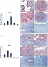
Figure 1
Figure 1. Expression of Msx1 and Msx2 in the uterus during early pregnancy.
Real-time PCR was performed to monitor the expression of mRNAs corresponding to Msx1 and Msx2 in uterus on days 1 to 5 of gestation. The relative levels of gene expression on different days of pregnancy were determined by setting the expression level of Msx1 mRNA (A, Left panel) and Msx2 mRNA (B, Left panel) on day 1 of pregnancy at 1.0. Rplp0, encoding a ribosomal protein, was used to normalize the level of RNA. Uterine sections from day 1 to day 5 (a–e) of pregnancy were subjected to immunohistochemical analysis using anti-MSX1 (A, Right panel) and anti-MSX2 (B, Right panel) antibodies. Panel f shows uterine sections from day 3 pregnant mice treated with non-immune IgG. L, G and S indicate luminal epithelium, glandular epithelium and stroma, respectively.
Figure 2
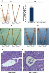
Figure 2. Lack of uterine Msx1 and Msx2 causes implantation failure.
A. Embryo implantation sites were examined in Msx1f/fMsx2f/f and Msx1d/dMsx2d/d mice by the vascular permeability assay, which can be scored as distinct blue bands (red arrows) following an injection of Chicago blue dye on day 5 of pregnancy (D5, n = 6) or direct eye-visualization of implanted embryo on day 6 (D6, n = 4) and on day 7 (D7, n = 4) of pregnancy. The graph represents the quantification of implantation sites in Msx1f/fMsx2f/f and Msx1d/dMsx2d/d mice on day 5 of pregnancy. B. Failure of embryo attachment in Msx1d/dMsx2d/d uteri. Histological analysis of uterine sections obtained from Msx1f/fMsx2f/f (a) and Msx1d/dMsx2d/d (b) mice on day 5 (n = 3) of pregnancy by Hematoxylin and Eosin staining. Note the intimate contact between embryo and luminal epithelium in Msx1f/fMsx2f/f mice and the free floating embryo in the uterine lumen of Msx1d/dMsx2d/d mice. L and E indicate luminal epithelium and embryo respectively.
Figure 3
Figure 3. Enhanced ESR1 activity in the luminal epithelium of Msx1d/dMsx2d/d uteri.
A. Uterine sections obtained from Msx1f/f Msx2f/f (left panel) and Msx1d/dMsx2d/d (right panel) mice on day 4 of pregnancy were subjected to IHC using antibodies against PGR (top panel, a and b), ESR1 (middle panel, c and d) and phospho-ESR1 (lower panel, e and f). B. Real-time PCR was performed to analyze the expression of E-regulated genes, lactotransferrin (Ltf), Clca3, lipocalin2 and Muc-1 in uteri of Msx1f/fMsx2f/f and Msx1d/dMsx2d/d mice on day 4 of pregnancy. The level of Ck18 was used as internal control to normalize gene expression. The data are represented as the mean fold induction ± SEM, *p<0.05. C. Real-time PCR was performed to analyze the expression of P-regulated genes, Ihh, COUP-TF II, Hand2 and Hoxa10, in uteri of Msx1f/fMsx2f/f and Msx1d/dMsx2d/d mice on day 4 of pregnancy. The level of Rplp0 or Ck18 was used as internal control to normalize gene expression.
Figure 4
Figure 4. Enhanced proliferation in the uterine epithelium and lack of receptivity in Msx1d/dMsx2d/d mice.
A. Immunohsitochemical localization of Ki67 in the uterine sections of Msx1f/fMsx2f/f (left panel, a and c) and Msx1d/dMsx2d/d (right panel, b and d) mice on day 4 of pregnancy. Panels a and b indicate lower magnification (20×) and c and d indicate higher magnification (40×). L and G indicate luminal epithelium and glandular epithelium respectively. B. Real-time PCR was performed to analyze the expression of glandular factors, Lif, Foxa2 and Spink3 in uteri of Msx1f/fMsx2f/f and Msx1d/dMsx2d/d mice on day 4 of pregnancy. The level of Ck18 was used as internal control to normalize gene expression. The data are represented as the mean fold induction ± SEM, ***p<0.0001. C. Transmission electron microscopy of uterine sections obtained from Msx1f/f Msx2f/f (left panel, a and b) and Msx1d/dMsx2d/d (right panel, c and d) mice on day 4 of pregnancy. Panels a and c indicate lower magnification (5Kx) and b and d indicate higher magnification (30Kx). D. Immunohistochemical analysis of Muc-1 expression in the uterine sections of Msx1f/fMsx2f/f (upper panel) and Msx1d/dMsx2d/d (lower panel) mice on day 1 (a and d), day 4 (b and e) and day 5 (c and f) of pregnancy. L indicates luminal epithelium.
Figure 5
Figure 5. Wnt/β-catenin signaling controls FGF synthesis in uterine stromal cells.
A. Real-time PCR was performed to analyze the expression of Wnt ligands in uterine epithelial cells of Msx1f/fMsx2f/f and Msx1d/dMsx2d/d mice on day 4 of pregnancy. The level of Ck18 was used as internal control to normalize gene expression. The data are represented as the mean fold induction ± SEM, *p<0.01, ***p<0.0001. B. Real-time PCR was performed to analyze the expression of Wnt ligands in uterine stromal cells of Msx1f/fMsx2f/f and Msx1d/dMsx2d/d mice on day 4 of pregnancy. C. Real-time PCR was performed to analyze the expression of Fgf family members in uterine stromal cells of Msx1f/fMsx2f/f and Msx1d/dMsx2d/d mice on day 4 of pregnancy. The level of Rplp0 was used as internal control to normalize gene expression. The data are represented as the mean fold induction ± SEM, *p<0.01, **p<0.001, ***p<0.0001. D. The level of active β-catenin in uterine sections of Msx1f/fMsx2f/f (left panel) and Msx1d/dMsx2d/d (right panel) mice on day 4 of pregnancy was analyzed by IHC. (Magnification: a and c: 10×, b and d: 40×) E. Primary stromal cells were isolated from uteri of Msx1d/dMsx2d/d mice on day 3 of pregnancy and transfected with siRNA targeted to the β-catenin mRNA. Total RNA was isolated 24 h after transfection to analyze the expression of Fgf family members by Real-time PCR. The level of Rplp0 was used as an internal control to normalize gene expression. The data are represented as the mean fold induction ± SEM, *p<0.01, **p<0.001, ***p<0.0001.
Figure 6
Figure 6. Enhanced FGFR signaling in the epithelium of Msx1d/dMsx2d/d uteri.
A. The level of p-FRS2 was examined in the uterine sections of Msx1f/fMsx2f/f (upper panel) and Msx1d/dMsx2d/d (lower panel) mice on day 4 of pregnancy by immunohistochemistry. Magnification: a and d: 10×, b and e: 20×, c and f: 40×. B. The level of p-ERK was examined in the uterine sections of Msx1f/fMsx2f/f (upper panel) and Msx1d/dMsx2d/d (lower panel) mice on day 4 of pregnancy by immunohistochemistry. Magnification: a and d: 10×, b and e: 20×, c and f: 40×. L, G and S indicate luminal epithelium, glandular epithelium, and stroma respectively. C. FGFR-specific inhibitor PD173074 was applied to one uterine horn of Msx1d/dMsx2d/d (n = 3) mice on day 3 of pregnancy. The other horn served as vehicle-treated control. Uterine horns were collected on day 4 morning and sections were subjected to immunohistochemistry to detect p-FRS2, Ki67, and Muc-1.
Figure 7
Figure 7. Mechanism of Msx1 and Msx2 action in mouse uterus.
In normal pregnancy, MSX1 and MSX2 act to repress WNT and β-catenin signaling and inhibit FGF synthesis in the uterine stroma, thereby suppressing stromal-epithelial cross-talk. In the absence of MSX1 and MSX2, FGFs are induced, activating the epithelial FGFR-ERK1/2 pathway, and promoting epithelial proliferation. Activated ERK1/2 then phosphorylates epithelial ESR1. This triggers transcriptional activation of ESR1 and expression of its target genes, such as Muc-1, which prevent the functional transformation of the luminal epithelium to receptive state, blocking embryo implantation.

Similar articles

Cited by

References

    1. Carson DD, Bagchi I, Dey SK, Enders AC, Fazleabas AT, et al. Embryo Implantation. Developmental Biology. 2000;223:217–237. - PubMed
    1. Dey SK, Lim H, Das SK, Reese J, Paria BC, et al. Molecular Cues to Implantation. Endocr Rev. 2004;25:341–373. - PubMed
    1. Ramathal CY, Bagchi IC, Taylor RN, Bagchi MK. Endometrial Decidualization: Of Mice and Men. Semin Reprod Med. 2010;28:17–26. - PMC - PubMed
    1. Rubel CA, Jeong J-W, Tsai SY, Lydon JP, DeMayo FJ. Epithelial-Stromal Interaction and Progesterone Receptors in the Mouse Uterus. Semin Reprod Med. 2010;28:027–035. - PubMed
    1. Lee KY, Jeong J-W, Tsai SY, Lydon JP, DeMayo FJ. Mouse models of implantation. Trends in Endocrinology & Metabolism. 2007;18:234–239. - PubMed

Publication types