. 2012;8(2):e1002527.
doi: 10.1371/journal.pgen.1002527. Epub 2012 Feb 23.

Sequestration of highly expressed mRNAs in cytoplasmic granules, P-bodies, and stress granules enhances cell viability

Affiliations

Sequestration of highly expressed mRNAs in cytoplasmic granules, P-bodies, and stress granules enhances cell viability

Anna Lavut et al. PLoS Genet. 2012.

Abstract

Transcriptome analyses indicate that a core 10%-15% of the yeast genome is modulated by a variety of different stresses. However, not all the induced genes undergo translation, and null mutants of many induced genes do not show elevated sensitivity to the particular stress. Elucidation of the RNA lifecycle reveals accumulation of non-translating mRNAs in cytoplasmic granules, P-bodies, and stress granules for future regulation. P-bodies contain enzymes for mRNA degradation; under stress conditions mRNAs may be transferred to stress granules for storage and return to translation. Protein degradation by the ubiquitin-proteasome system is elevated by stress; and here we analyzed the steady state levels, decay, and subcellular localization of the mRNA of the gene encoding the F-box protein, UFO1, that is induced by stress. Using the MS2L mRNA reporter system UFO1 mRNA was observed in granules that colocalized with P-bodies and stress granules. These P-bodies stored diverse mRNAs. Granules of two mRNAs transported prior to translation, ASH1-MS2L and OXA1-MS2L, docked with P-bodies. HSP12 mRNA that gave rise to highly elevated protein levels was not observed in granules under these stress conditions. ecd3, pat1 double mutants that are defective in P-body formation were sensitive to mRNAs expressed ectopically from strong promoters. These highly expressed mRNAs showed elevated translation compared with wild-type cells, and the viability of the mutants was strongly reduced. ecd3, pat1 mutants also exhibited increased sensitivity to different stresses. Our interpretation is that sequestration of highly expressed mRNAs in P-bodies is essential for viability. Storage of mRNAs for future regulation may contribute to the discrepancy between the steady state levels of many stress-induced mRNAs and their proteins. Sorting of mRNAs for future translation or decay by individual cells could generate potentially different phenotypes in a genetically identical population and enhance its ability to withstand stress.


Conflict of interest statement

The authors have declared that no competing interests exist.

Figures

Figure 1
Figure 1. Transcription of UFO1 in response to arsenate, H2O2, and UV.
A. Wild type cells were grown in SC 2% glucose medium overnight, diluted to A 600 = 0.1, regrown to A 600 = 0.5 and treated with 1 mM arsenate, 8.8 mM H2O2, or irradiated with 40 mJ/cm2 UV. Aliquots were collected at the indicated times and analyzed by qRT-PCR. mRNA levels were normalized to ACT1 and to time 0 (untreated log cells). B. pGAL-GFP-UFO1 was expressed in ufo1Δ mutants by overnight induction with 2% galactose. Next morning cells were diluted to A 600 = 0.1, regrown to A 600 = 0.5 then untreated, or stressed with 1 mM arsenate or 8.8 mM H2O2 for 30 minutes, or irradiated with 40 mJ/cm2 UV. The cells were washed and transferred to SC with 4% glucose. Samples were collected immediately after addition of glucose and at the times indicated and analyzed by qRT-PCR. mRNA levels were normalized to ACT1 and to time 0 (untreated log cells). C. Western blot analysis of Ufo1GFP protein produced from the tagged genomic UFO1-GFP gene. Anti-GFP antibodies were used to detect Ufo1GFP and anti-α-tubulin antibodies to detect α-tubulin that serves as a loading control. D. UFO1 mRNA levels in untreated wild type, yap1Δ or pdr1Δ mutants. mRNA levels were normalized to ACT1 and to w.t. mRNA levels. E. yap1Δ or pdr1Δ mutants grown, treated and analyzed as in Figure 1A. mRNA levels were normalized to ACT1 and to time 0 (untreated log cells).
Figure 2
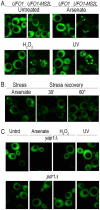
Figure 2. Stress-induced granules appear only in UFO1-MS2L stressed cells.
A. Wild type and UFO1-MS2L cells transformed with pCP-MS2L-GFPx3 at A 600 = 0.5, were transferred to SC 2% glucose without methionine for 1 hour to induce the CPGFP. Cells were untreated or exposed to 1 mM arsenate, 8.8 mM H2O2, or UV-irradiated with 40 mJ/cm2. Aliquots were collected 30 minutes after each treatment. Fluorescent granules appear only in cells with the tagged UFO1-MS2L gene. B. UFO1-MS2L cells expressing pCP-MS2L-GFPx3 grown as above, treated with 1 mM arsenate for 30 minutes (stress), and transferred to fresh SC glucose medium. Stress recovery 30′ and 60′ indicate time after transfer to fresh SC. C. UFO1-MS2L, yap1Δ or UFO1-MS2L, pdr1Δ cells grown as above and untreated (Utrd), exposed to 1 mM arsenate, 8.8 mM H2O2, or UV-irradiated with 40 mJ/cm2.
Figure 3
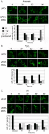
Figure 3. Time course of granule appearance in UFO1-MS2L cells after stress.
Cells treated with A. 1 mM arsenate, B. 8.8 mM H2O2, or C. 40 mJ/cm2 UV, and visualized by confocal microscopy at the times indicated. Histograms show number of cells with different numbers of granules per cell at the times indicated (n = >150 cells).
Figure 4
Figure 4. UFO1-MS2L mRNAs induced by stress colocalize with subunits of PBs and SGs.
A. UFO1-MS2L cells at A 600 = 0.5 that produce PB marker protein, Dcp1RFP, or the SG marker, eIF4ERFP, were transferred to fresh SC 2% glucose medium without methionine for 1 hour. B. Cells as in A, deprived for glucose for 30 minutes. C. Cells as in B, treated for 30 minutes with 1 mM arsenate, or D. 8.8 mM H2O2. E. Cells at A 600 = 0.5 that produce both the PB marker protein, Dhh1GFP, and eIF4ERFP, treated with 1 mM arsenate for 30 minutes in the presence or the absence of glucose.
Figure 5
Figure 5. UFO1-MS2L and MFA2-U1A mRNAs are sequestered in the same PBs after arsenate stress.
A. Control wild type cells at A 600 = 0.5 producing U1AGFP untreated or treated with 1 mM arsenate for 30 minutes. B. wild type UFO1-MS2L cells at A 600 = 0.5 expressing pMFA2-U1A, with their respective RNA-binding proteins, CPmCherry and U1AGFP, untreated or treated with 1 mM arsenate and stained with Hoechst 33342 at a final concentration of 2.5 µg/mL for 30 minutes.
Figure 6
Figure 6. ASH1 and OXA1 mRNA granules interact with PBs.
A. ASH1-MS2L cells at A 600 = 0.5 with pCP-MS2L-GFPx3 and the PB marker, Edc3mCherry, either untreated, treated with 1 mM arsenate or with 8.8 mM H2O2 for 30 minutes or transferred to SC without glucose for 30 minutes. Merge ×5 represents 5 times enlargement of selected granules indicated with white arrows in the whole cells. B. OXA1-MS2L cells treated as in A., and visualized by confocal microscopy. C. Histograms of ASH1-MS2L cells or D. OXA1-MS2L cells, showing percentages of overlapping, docked, or distinct granule types in a population of cells untreated, treated with 1 mM arsenate or with 8.8 mM H2O2 for 30 minutes or stressed in SC without glucose (n = >100 cells).
Figure 7
Figure 7. Induction of Hsp12 protein and mRNA, and mRNA decay after stress.
A. WB of protein produced from genomic HSP12-GFP in response to stress. B. UFO1 and HSP12 mRNA levels in untreated cells analyzed by qRT-PCR. mRNA levels were normalized to ACT1. C. Induction of HSP12 mRNA by stress. Wild type cells at A 600 = 0.5, treated with 1 mM arsenate, 8.8 mM H2O2, irradiated with 40 mJ/cm2 UV or shifted from 30°C to 37°C for 40 minutes. Aliquots were collected at the times indicated and analyzed by qRT-PCR. D. HSP12 mRNA decay. pGAL-HSP12 was expressed in hsp12Δ mutants by overnight induction with 2% galactose. Next morning cells at A 600 = 0.5 were untreated, or stressed with 1 mM arsenate or 8.8 mM H2O2 for 30 minutes, or irradiated with 40 mJ/cm2 UV. The cells were washed and transferred to SC medium with 4% glucose. Samples were collected immediately after addition of glucose and at the times indicated and analyzed by qRT-PCR. mRNA levels were normalized to ACT1 and to time 0 (untreated log cells).
Figure 8
Figure 8. Ectopic high level gene expression affects viability of mutants unable to form PBs and SGs.
A. Visualization of wild type, pat1Δ, edc3Δ, pat1Δ or edc3Δ, lsm4Δc cells expressing the PB marker Dcp2mCherry and the SG marker Pab1GFP untreated or exposed for 30 minutes to 1 mM arsenate or 8.8 mM H2O2, irradiated with 40 mJ/cm2 UV, or incubated in SC medium without glucose for 30 minutes. B. Viability of wild type, pat1Δ; edc3Δ, pat1Δ or edc3Δ, lsm4Δc cells untreated (Unt) or treated with 1 mM arsenate, 8.8 mM H2O2 or irradiated with 40 mJ/cm2 UV analyzed by the spot test viability assay. C. Wild type, pat1Δ; edc3Δ, pat1Δ or edc3Δ, lsm4Δc cells expressing empty pGAL-vector (YCp), pGAL-GFP-UFO1, pGAL-HSP12 or MFA2-U1A (pRP1193) were grown in SC medium with 2% glucose or induced in 2% galactose medium overnight, diluted and regrown in the same media to A 600 = 0.5 for spot test analysis on SC plates with 2% glucose or 2% galactose, respectively. D. WB analysis of w.t or edc3Δ, pat1Δ cells expressing UFO1, HSP12 or MFA2 from the GAL promoter. Glu (noninducing conditions) and Gal (inducing). The intensities of each protein band were normalized to the α-tubulin loading control using ImageJ . E. Comparison of UFO1 and HSP12 mRNA decay. pGAL-GFP-UFO1 or pGAL-HSP12 was expressed in w.t or edc3Δ, pat1Δ cells by overnight induction with 2% galactose. Next morning cells at A 600 = 0.5 were washed and transferred to SC medium with 4% glucose. Samples were collected immediately after addition of glucose and at the times indicated and analyzed by qRT-PCR. mRNA levels were normalized to ACT1 and to time 0 (untreated log cells).
Figure 9
Figure 9. Pathways for mRNA under normal and stress conditions.
Under normal growth conditions after transcription a mRNA could go directly to the polysomes for translation (a), or the mRNAs could be escorted by subunits of the RNA polymerase complex to the PBs and sorted either for the polysomes for translation (b), or the mRNA could undergo decay in the PBs (c). Under stress conditions pre-existing mRNAs could undergo enhanced translation as we propose for HSP12 mRNA (α), or be retracted from the polysomes (β) for future sorting for storage or decay (γ or δ). Furthermore, after stress mRNAs can shuttle between PBs and SGs (γ) from where they can return to translation (ε). mRNA lifecycles under normal growth conditions have blue arrows, stress conditions are indicated with brown arrows. A more comprehensive description of the mRNA lifecycle, particularly of mRNA decay, can be found in .

Similar articles

Cited by

References

    1. Gasch A, Werner-Washburne M. The genomics of yeast responses to environmental stress and starvation. Funct Integr Genomics. 2002;2:181–192. - PubMed
    1. Gasch AP, Huang M, Metzner S, Botstein D, Elledge SJ, et al. Genomic expression responses to DNA-damaging agents and the regulatory role of the yeast ATR homolog Mec1p. Mol Biol Cell. 2001;12:2987–3003. - PMC - PubMed
    1. Temple MD, Perrone GG, Dawes IW. Complex cellular responses to reactive oxygen species. Trends Cell Biol. 2005;15:319–326. - PubMed
    1. Gasch AP, Spellman PT, Kao CM, Carmel-Harel O, Eisen MB, et al. Genomic expression programs in the response of yeast cells to environmental changes. Mol Biol Cell. 2000;11:4241–4257. - PMC - PubMed
    1. Hershko A, Ciechanover A. The ubiquitin system. Annu Rev Biochem. 1998;67:425–479. - PubMed

Publication types

MeSH terms