

doi: 10.4021/jocmr661w.
Epub 2011 Sep 26.
Images of cecal volvulus from a strangulating fallopian tube: a case report
Affiliations
- PMID: 22383914
- PMCID: PMC3279488
- DOI: 10.4021/jocmr661w
Item in Clipboard
Images of cecal volvulus from a strangulating fallopian tube: a case report
J Clin Med Res.
2011 Oct.
Abstract
An unusual case of cecal volvulus arising from a strangulating fallopian tube is presented. The etiology, diagnosis, and management guidelines of this infrequent cause of large bowel obstruction are reviewed. Computed tomography images are included, which demonstrate key features that are pathognomonic for this condition. To our knowledge, this is the first report of gynecologic adnexa giving rise to cecal volvulus.
Keywords: Cecal volvulus; Gynecologic and general surgery; Intestinal obstruction.
Figures
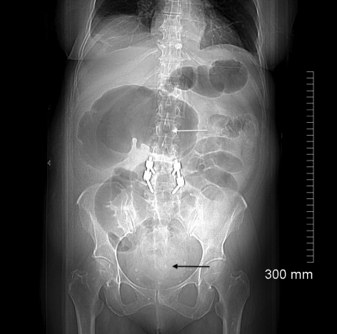
CT scout image demonstrates a dilated cecum in the right upper quadrant producing the classic ”coffee bean“ sign (white arrow). There are multiple dilated loops of small bowel present centrally within the abdomen with a paucity of gas in the distal colon (black arrow) suggestive of a complete intestinal obstruction. This patient had spinal instrumentation consistent with her previous history of spine surgery.
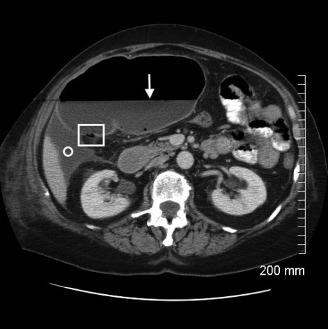
Axial CT image with intravenous and oral contrast through the level of the mid-abdomen demonstrates massive dilatation of the cecum. There is a prominent air-fluid level (white arrow) and multiple locules of air are noted in the dependent portion of the wall consistent with pneumatosis intestinalis (square). However, no extraluminal free air is present to suggest frank perforation and the wall of the cecum continues to enhance uniformly. The associated perihepatic free fluid (circle) is consistent with a high-grade type of intestinal obstruction.
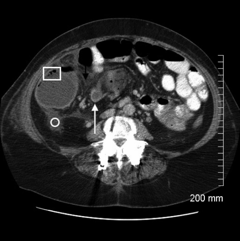
Axial CT image demonstrates twisting of the mesenteric vessels producing the ”CT whirl“ sign, which is highly specific for intestinal volvulus (white arrow). Again demonstrated are the associated pneumatosis intestinalis (square) and pericolonic free fluid (circle).
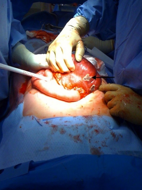
Laparotomy revealed a massively dilated cecum in the right upper quadrant consistent with radiological findings. Turbid free fluid was encountered upon entry into the abdomen. The cecum demonstrated patchy areas of ischemic necrosis (black arrow) and despite carefully twisting it free from its strangulated mesentery, remained unviable. A right hemicolectomy with primary ileocolic anastomosis was performed.
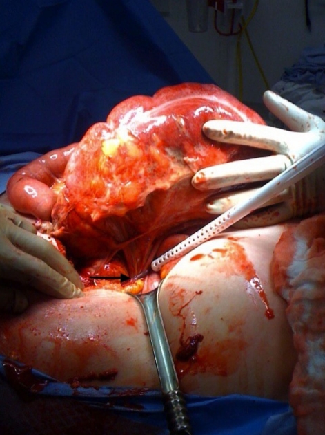
A tubular structure was found to be strangulating the cecal mesentery (black arrow). This tubular structure was gently dissected free and found to originate in the pelvis. The patient had a remote history of abdominal hysterectomy and it was unclear whether gynecological adnexae were concurrently removed. An intra-operative gynecological consult was obtained and the tubular structure was identified as the remnant right fallopian tube and confirmed on pathology. A bilateral salping-oophorectomy was performed.
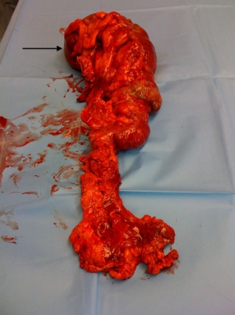
Right hemicolectomy specimen demonstrating areas of intestinal gangrene from closed loop obstruction and ischemic necrosis (black arrow indicates cecum). Pathological examination confirmed the presence of full-thickness intestinal wall necrosis. There were no intraluminal masses identified in the pathology specimen.
References
-
- Habre J, Sautot-Vial N, Marcotte C, Benchimol D. Caecal volvulus. Am J Surg. 2008;196(5):e48–49. - PubMed
-
- Mathew HM, Chin K. Caecal volvulus--an unusual cause of post-operative pain following gynaecological surgery. J Obstet Gynaecol. 2003;23(6):685–686. - PubMed
-
- Moore CJ, Corl FM, Fishman EK. CT of cecal volvulus: unraveling the image. AJR Am J Roentgenol. 2001;177(1):95–98. - PubMed
-
- Schaeffer AJ, Hines JR. Cecal volvulus after obstetric and gynecologic procedures. Report of 3 patients. Obstet Gynecol. 1971;37(2):255–259. - PubMed
LinkOut - more resources
Full Text Sources