

LEM-3 - A LEM domain containing nuclease involved in the DNA damage response in C. elegans
- PMID: 22383942
- PMCID: PMC3285610
- DOI: 10.1371/journal.pone.0024555
LEM-3 - A LEM domain containing nuclease involved in the DNA damage response in C. elegans
Abstract
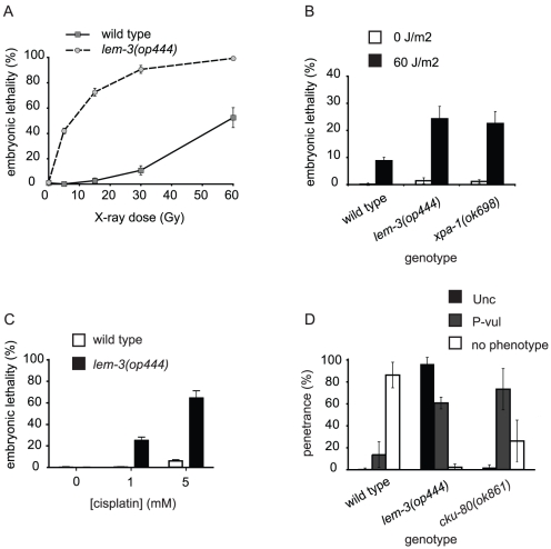
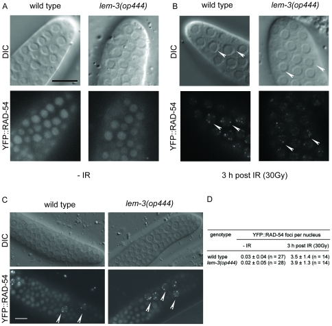
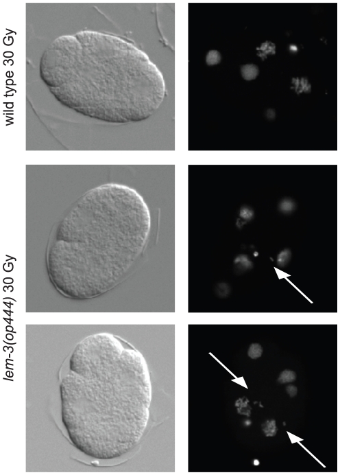
The small nematode Caenorhabditis elegans displays a spectrum of DNA damage responses similar to humans. In order to identify new DNA damage response genes, we isolated in a forward genetic screen 14 new mutations conferring hypersensitivity to ionizing radiation. We present here our characterization of lem-3, one of the genes identified in this screen. LEM-3 contains a LEM domain and a GIY nuclease domain. We confirm that LEM-3 has DNase activity in vitro. lem-3(lf) mutants are hypersensitive to various types of DNA damage, including ionizing radiation, UV-C light and crosslinking agents. Embryos from irradiated lem-3 hermaphrodites displayed severe defects during cell division, including chromosome mis-segregation and anaphase bridges. The mitotic defects observed in irradiated lem-3 mutant embryos are similar to those found in baf-1 (barrier-to-autointegration factor) mutants. The baf-1 gene codes for an essential and highly conserved protein known to interact with the other two C. elegans LEM domain proteins, LEM-2 and EMR-1. We show that baf-1, lem-2, and emr-1 mutants are also hypersensitive to DNA damage and that loss of lem-3 sensitizes baf-1 mutants even in the absence of DNA damage. Our data suggest that BAF-1, together with the LEM domain proteins, plays an important role following DNA damage - possibly by promoting the reorganization of damaged chromatin.
Conflict of interest statement
Figures

Similar articles
-
The conserved LEM-3/Ankle1 nuclease is involved in the combinatorial regulation of meiotic recombination repair and chromosome segregation in Caenorhabditis elegans.PLoS Genet. 2018 Jun 7;14(6):e1007453. doi: 10.1371/journal.pgen.1007453. eCollection 2018 Jun. PLoS Genet. 2018. PMID: 29879106 Free PMC article.
-
Caenorhabditis elegans BUB-3 and SAN-1/MAD3 Spindle Assembly Checkpoint Components Are Required for Genome Stability in Response to Treatment with Ionizing Radiation.G3 (Bethesda). 2017 Dec 4;7(12):3875-3885. doi: 10.1534/g3.117.1122. G3 (Bethesda). 2017. PMID: 29046436 Free PMC article.
-
Functional dissection of the conserved C. elegans LEM-3/ANKLE1 nuclease reveals a crucial requirement for the LEM-like and GIY-YIG domains for DNA bridge processing.Nucleic Acids Res. 2025 Mar 20;53(6):gkaf265. doi: 10.1093/nar/gkaf265. Nucleic Acids Res. 2025. PMID: 40193711 Free PMC article.
-
[Structure and biological functions of mammalian LEM-domain proteins].Yi Chuan. 2015 Feb;37(2):128-139. doi: 10.16288/j.yczz.14-351. Yi Chuan. 2015. PMID: 25665639 Review. Chinese.
-
Lamina-associated polypeptide (LAP)2α and other LEM proteins in cancer biology.Adv Exp Med Biol. 2014;773:143-63. doi: 10.1007/978-1-4899-8032-8_7. Adv Exp Med Biol. 2014. PMID: 24563347 Free PMC article. Review.
Cited by
-
Familial partial lipodystrophy, mandibuloacral dysplasia and restrictive dermopathy feature barrier-to-autointegration factor (BAF) nuclear redistribution.Cell Cycle. 2012 Oct 1;11(19):3568-77. doi: 10.4161/cc.21869. Epub 2012 Aug 30. Cell Cycle. 2012. PMID: 22935701 Free PMC article.
-
Nuclear envelope assembly relies on CHMP-7 in the absence of BAF-LEM-mediated hole closure.bioRxiv [Preprint]. 2023 Jul 7:2023.07.06.547980. doi: 10.1101/2023.07.06.547980. bioRxiv. 2023. Update in: J Cell Sci. 2023 Nov 1;136(21):jcs261385. doi: 10.1242/jcs.261385. PMID: 37461528 Free PMC article. Updated. Preprint.
-
Multilayered Reprogramming in Response to Persistent DNA Damage in C. elegans.Cell Rep. 2017 Aug 29;20(9):2026-2043. doi: 10.1016/j.celrep.2017.08.028. Cell Rep. 2017. PMID: 28854356 Free PMC article.
-
Formation and resolution of meiotic chromosome entanglements and interlocks.J Cell Sci. 2024 Jul 1;137(13):jcs262004. doi: 10.1242/jcs.262004. Epub 2024 Jul 10. J Cell Sci. 2024. PMID: 38985540 Free PMC article. Review.
-
Unique and shared functions of nuclear lamina LEM domain proteins in Drosophila.Genetics. 2014 Jun;197(2):653-65. doi: 10.1534/genetics.114.162941. Epub 2014 Apr 3. Genetics. 2014. PMID: 24700158 Free PMC article.
References
-
- Hoeijmakers JHJ. DNA Damage, Aging, and Cancer. N Engl J Med. 2009;361:1475–1485. doi: 10.1056/NEJMra0804615. - DOI - PubMed
-
- Stergiou L, Hengartner MO. Death and more: DNA damage response pathways in the nematode C. elegans. Cell Death Differ. 2004;11:21–28. doi: 10.1038/sj.cdd.4401340. - DOI - PubMed
-
- O'Neil N, Rose A. DNA repair. Worm Book. 2006:1–12. doi: 10.1895/wormbook.1.54.1. - DOI - PMC - PubMed
-
- Gartner A, Boag PR, Blackwell TK. Germline survival and apoptosis. Worm Book. 2008:1–20. doi: 10.1895/wormbook.1.145.1. - DOI - PMC - PubMed
-
- Gartner A, Milstein S, Ahmed S, Hodgkin J, Hengartner MO. A conserved checkpoint pathway mediates DNA damage–induced apoptosis and cell cycle arrest in C. elegans. Mol Cell. 2000;5:435–443. - PubMed
Publication types
MeSH terms
Substances
LinkOut - more resources
Full Text Sources
Molecular Biology Databases
Research Materials
