

A nonluminescent and highly virulent Vibrio harveyi strain is associated with "bacterial white tail disease" of Litopenaeus vannamei shrimp
- PMID: 22383954
- PMCID: PMC3288001
- DOI: 10.1371/journal.pone.0029961
A nonluminescent and highly virulent Vibrio harveyi strain is associated with "bacterial white tail disease" of Litopenaeus vannamei shrimp
Abstract
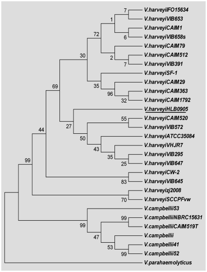
Recurrent outbreaks of a disease in pond-cultured juvenile and subadult Litopenaeus vannamei shrimp in several districts in China remain an important problem in recent years. The disease was characterized by "white tail" and generally accompanied by mass mortalities. Based on data from the microscopical analyses, PCR detection and 16S rRNA sequencing, a new Vibrio harveyi strain (designated as strain HLB0905) was identified as the etiologic pathogen. The bacterial isolation and challenge tests demonstrated that the HLB0905 strain was nonluminescent but highly virulent. It could cause mass mortality in affected shrimp during a short time period with a low dose of infection. Meanwhile, the histopathological and electron microscopical analysis both showed that the HLB0905 strain could cause severe fiber cell damages and striated muscle necrosis by accumulating in the tail muscle of L. vannamei shrimp, which led the affected shrimp to exhibit white or opaque lesions in the tail. The typical sign was closely similar to that caused by infectious myonecrosis (IMN), white tail disease (WTD) or penaeid white tail disease (PWTD). To differentiate from such diseases as with a sign of "white tail" but of non-bacterial origin, the present disease was named as "bacterial white tail disease (BWTD)". Present study revealed that, just like IMN and WTD, BWTD could also cause mass mortalities in pond-cultured shrimp. These results suggested that some bacterial strains are changing themselves from secondary to primary pathogens by enhancing their virulence in current shrimp aquaculture system.
Conflict of interest statement
Figures


Similar articles
-
Integrating short- and full-length 16S rRNA gene sequencing to elucidate microbiome profiles in Pacific white shrimp (Litopenaeus vannamei) ponds.Microbiol Spectr. 2024 Nov 5;12(11):e0096524. doi: 10.1128/spectrum.00965-24. Epub 2024 Sep 27. Microbiol Spectr. 2024. PMID: 39329828 Free PMC article.
-
'Bright-red' syndrome in Pacific white shrimp Litopenaeus vannamei is caused by Vibrio harveyi.Dis Aquat Organ. 2010 Oct 26;92(1):11-9. doi: 10.3354/dao02274. Dis Aquat Organ. 2010. PMID: 21166310
-
Nitric oxide as an antimicrobial molecule against Vibrio harveyi infection in the hepatopancreas of Pacific white shrimp, Litopenaeus vannamei.Fish Shellfish Immunol. 2015 Jan;42(1):114-20. doi: 10.1016/j.fsi.2014.10.042. Epub 2014 Nov 7. Fish Shellfish Immunol. 2015. PMID: 25449376
-
White feces syndrome in shrimp: Comprehensive understanding of immune system responses.Fish Shellfish Immunol. 2024 Aug;151:109704. doi: 10.1016/j.fsi.2024.109704. Epub 2024 Jun 14. Fish Shellfish Immunol. 2024. PMID: 38880362 Review.
-
Vibrio harveyi: a serious pathogen of fish and invertebrates in mariculture.Mar Life Sci Technol. 2020;2(3):231-245. doi: 10.1007/s42995-020-00037-z. Epub 2020 Apr 3. Mar Life Sci Technol. 2020. PMID: 32419972 Free PMC article. Review.
Cited by
-
Evaluation of Therapeutic Efficiency of Stylicin against Vibrio parahaemolyticus Infection in Shrimp Penaeus vannamei through Comparative Proteomic Approach.Probiotics Antimicrob Proteins. 2024 Feb;16(1):76-92. doi: 10.1007/s12602-022-10006-w. Epub 2022 Dec 2. Probiotics Antimicrob Proteins. 2024. PMID: 36459385
-
Healthier Communities of Phytoplankton and Bacteria Achieved via the Application of Modified Clay in Shrimp Aquaculture Ponds.Int J Environ Res Public Health. 2021 Nov 4;18(21):11569. doi: 10.3390/ijerph182111569. Int J Environ Res Public Health. 2021. PMID: 34770083 Free PMC article.
-
Evidence of bacterioplankton community adaptation in response to long-term mariculture disturbance.Sci Rep. 2015 Oct 16;5:15274. doi: 10.1038/srep15274. Sci Rep. 2015. PMID: 26471739 Free PMC article.
-
Exosomal miRNAs in the plasma of Cynoglossus semilaevis infected with Vibrio harveyi: Pleiotropic regulators and potential biomarkers involved in inflammatory and immune responses.Front Immunol. 2022 Aug 18;13:949670. doi: 10.3389/fimmu.2022.949670. eCollection 2022. Front Immunol. 2022. PMID: 36059498 Free PMC article.
-
A metagenomic comparison of clearwater, probiotic, and Rapid BFTTM on Pacific whiteleg shrimp, Litopenaeus vannamei cultures.PeerJ. 2023 Sep 28;11:e15758. doi: 10.7717/peerj.15758. eCollection 2023. PeerJ. 2023. PMID: 37790619 Free PMC article.
References
-
- Qian D, Shi Z, Zhang S, Cao Z, Liu W, et al. Extra small virus-like particles (XSV) and nodavirus associated with whitish muscle disease in the giant freshwater prawn, Macrobrachium rosenbergii. J Fish Dis. 2003;26:521–527. - PubMed
-
- Lightner DV, Pantoja CR, Poulos BT, Tang KFJ, Redman RM, et al. Infectious myonecrosis: new disease in Pacific white shrimp. Glob Aquac Advocate. 2004;7:85.
-
- Tang KFJ, Pantoja CR, Redman RM, Lightner DV. Development if in situ hybridization and RT-PCR assay for the detection of a nodavirus (PvNV) that causes muscle necrosis in Penaeus vannamei. Dis Aquat Org. 2007;75:183–190. - PubMed
-
- Poulos BT, Lightner DV. Detection of infectious myonecrosis virus (IMNV) of penaeid shrimp by reverse-transcriptase polymerase chain reaction (RT-PCR). Dis Aquat Organ. 2006;73(1):69–72. - PubMed
-
- Lightner DV, Redman RM. Shrimp diseases and current diagnostic methods. Aquaculture. 1998;164:201–220.
Publication types
MeSH terms
Substances
LinkOut - more resources
Full Text Sources
Molecular Biology Databases
