. 2012;7(2):e30434.
doi: 10.1371/journal.pone.0030434. Epub 2012 Feb 24.

HemaMax™, a recombinant human interleukin-12, is a potent mitigator of acute radiation injury in mice and non-human primates

Affiliations

HemaMax™, a recombinant human interleukin-12, is a potent mitigator of acute radiation injury in mice and non-human primates

Lena A Basile et al. PLoS One. 2012.

Abstract

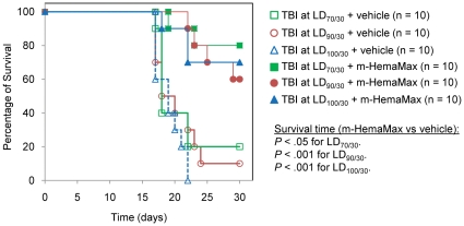
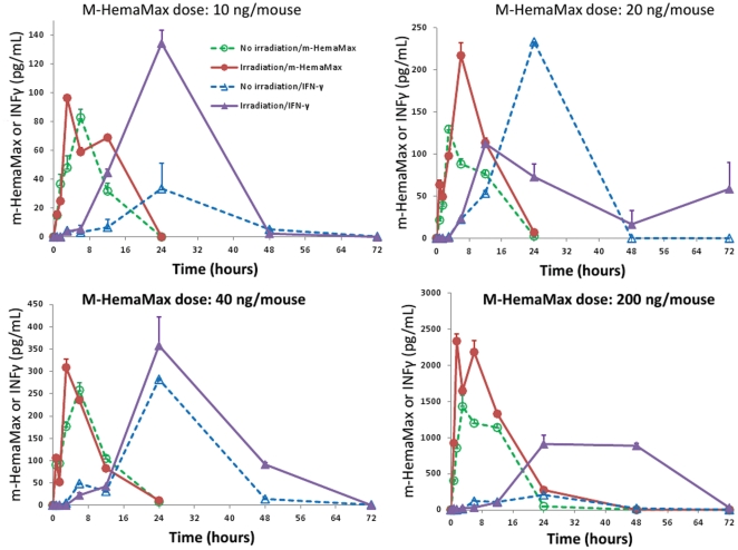
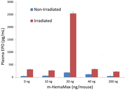
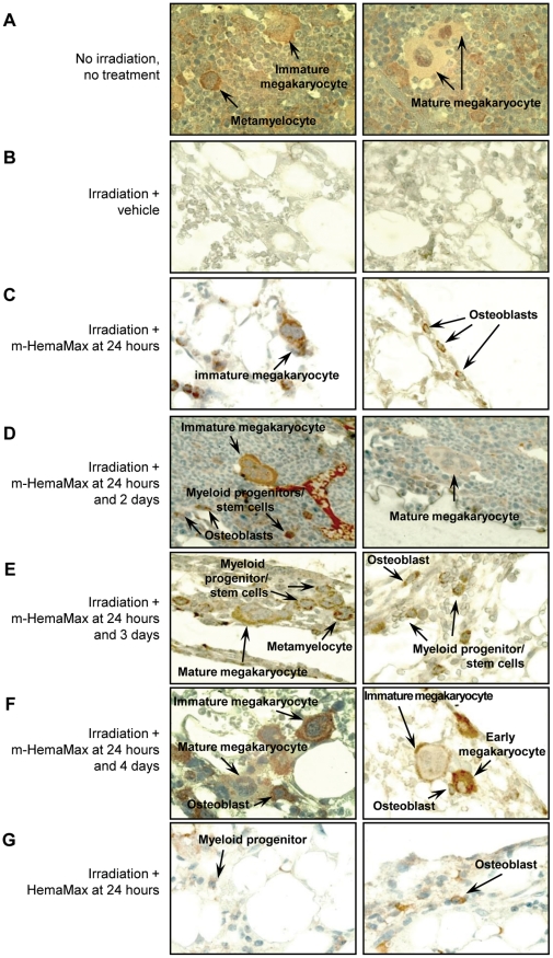
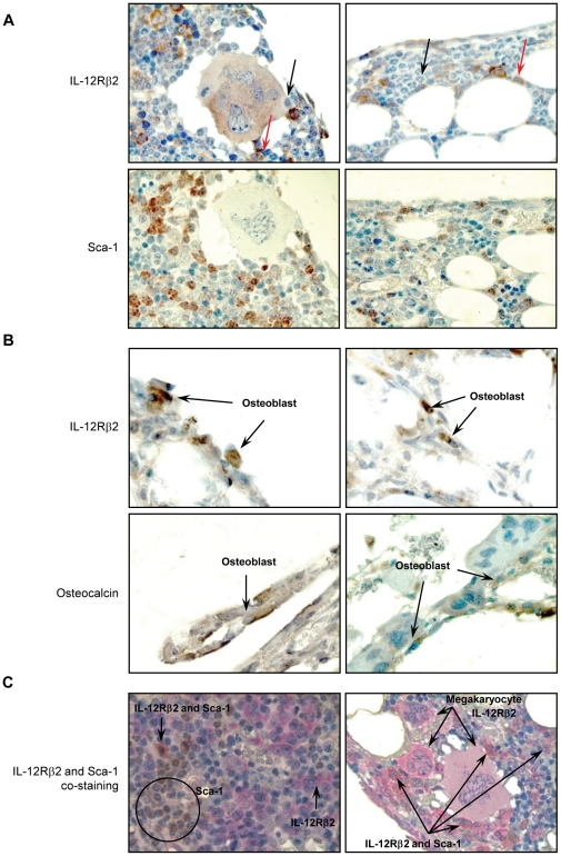
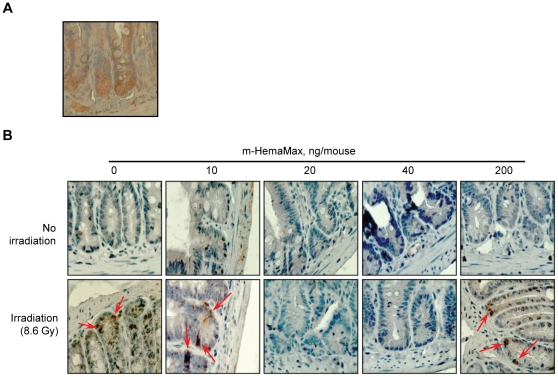
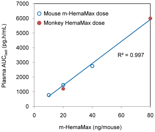
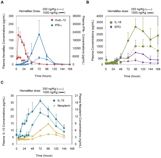
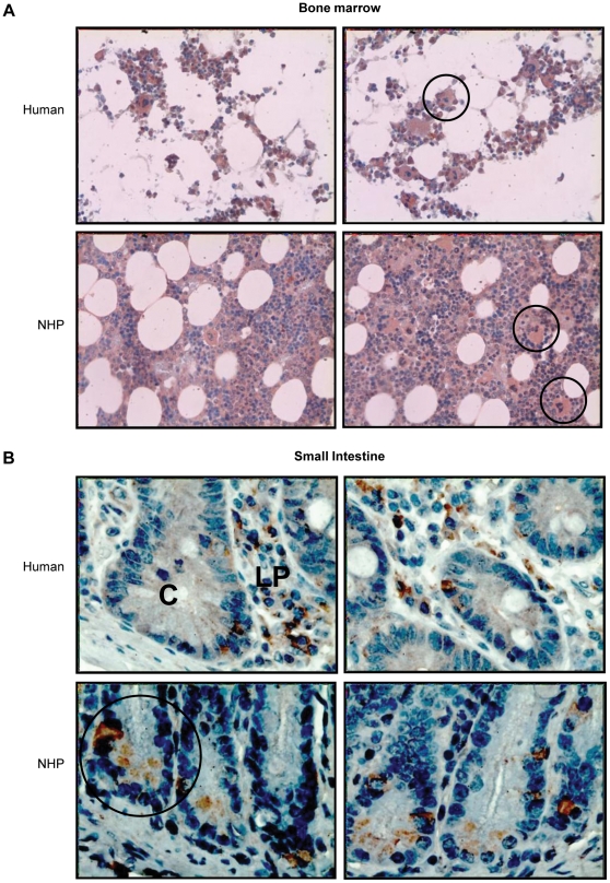
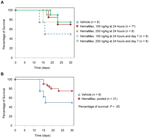
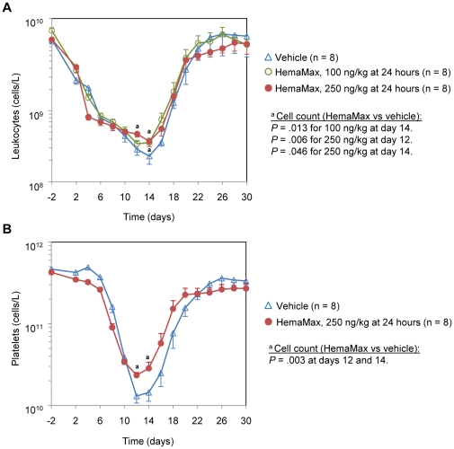
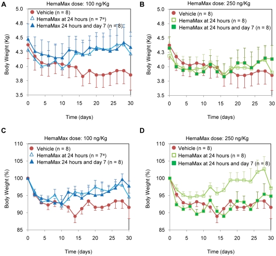
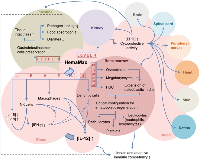
HemaMax, a recombinant human interleukin-12 (IL-12), is under development to address an unmet medical need for effective treatments against acute radiation syndrome due to radiological terrorism or accident when administered at least 24 hours after radiation exposure. This study investigated pharmacokinetics, pharmacodynamics, and efficacy of m-HemaMax (recombinant murine IL-12), and HemaMax to increase survival after total body irradiation (TBI) in mice and rhesus monkeys, respectively, with no supportive care. In mice, m-HemaMax at an optimal 20 ng/mouse dose significantly increased percent survival and survival time when administered 24 hours after TBI between 8-9 Gy (p<0.05 Pearson's chi-square test). This survival benefit was accompanied by increases in plasma interferon-γ (IFN-γ) and erythropoietin levels, recovery of femoral bone hematopoiesis characterized with the presence of IL-12 receptor β2 subunit-expressing myeloid progenitors, megakaryocytes, and osteoblasts. Mitigation of jejunal radiation damage was also examined. At allometrically equivalent doses, HemaMax showed similar pharmacokinetics in rhesus monkeys compared to m-HemaMax in mice, but more robustly increased plasma IFN-γ levels. HemaMax also increased plasma erythropoietin, IL-15, IL-18, and neopterin levels. At non-human primate doses pharmacologically equivalent to murine doses, HemaMax (100 ng/Kg and 250 ng/Kg) administered at 24 hours after TBI (6.7 Gy/LD(50/30)) significantly increased percent survival of HemaMax groups compared to vehicle (p<0.05 Pearson's chi-square test). This survival benefit was accompanied by a significantly higher leukocyte (neutrophils and lymphocytes), thrombocyte, and reticulocyte counts during nadir (days 12-14) and significantly less weight loss at day 12 compared to vehicle. These findings indicate successful interspecies dose conversion and provide proof of concept that HemaMax increases survival in irradiated rhesus monkeys by promoting hematopoiesis and recovery of immune functions and possibly gastrointestinal functions, likely through a network of interactions involving dendritic cells, osteoblasts, and soluble factors such as IL-12, IFN-γ, and cytoprotectant erythropoietin.


Conflict of interest statement

Competing Interests: Lena A. Basile, Dolph Ellefson, Zoya Gluzman-Poltorak, Katiana Junes-Gill, Vernon Mar, Sarita Mendonca, Jamie Tom, Alice Trinh, and Timothy K. Gallaher are employees of Neumedicines Inc. Joseph D. Miller is a consultant to Neumedicines Inc. HemaMax is a Neumedicines' product; there are numerous relevant patents, details of which are available on request. This does not alter the authors' adherence to all the PLoS ONE policies on sharing data and materials.

Figures

Figure 1
Figure 1. m-HemaMax administered at least 24 hours after TBI increased survival time of irradiated mice.
(a) Animals received vehicle or m-HemaMax at an ostensible dose of 100 ng/mouse at 24 hours and 72 hours after a TBI of 8 Gy (LD86/30). (b) Animals received vehicle or a single, ostensible dose of 300 ng/mouse of m-HemaMax at 24 hours, 48 hours, or 72 hours after a TBI of 9 Gy (LD100/30). (c) Animals received vehicle or a single low dose of m-HemaMax (2 ng/mouse or 18 ng/mouse) at 24 hours after a TBI of 7.9 Gy (LD85/30). Vehicle and m-HemaMax were injected subcutaneously. Vehicle was PBS in (a) and (b) and P5.6TT in (c). The delivered m-HemaMax dose was estimated to be 10 ng/mouse in (a) and 30 ng/mouse in (b) because subsequent studies showed that the actual m-HemaMax dose delivered was approximately 10% of the intended dose, most likely due to m-HemaMax sticking to surfaces of vials and syringes.
Figure 2
Figure 2. Efficacy of m-HemaMax in increasing survival is not dependent on radiation dose in mice.
Animals were subjected to TBI at ascending radiation doses of 8.6 Gy (LD70/30), 8.8 Gy (LD90/30), and 9.0 Gy (LD100/30) and subsequently received m-HemaMax at a dose of 20 ng/mouse 24 hours after irradiation. Mice were monitored for survival up to day 30. Vehicle was P5.6TT.
Figure 3
Figure 3. m-HemaMax administration increased plasma m-HemaMax and IFN-γ levels in irradiated and non-irradiated mice.
Animals received m-HemaMax subcutaneously at a dose of (a) 10 ng/mouse, (b) 20 ng/mouse, (c) 40 ng/mouse, or (d) 200 ng/mouse in the absence of irradiation or at 24 hours after an LD90/30 of TBI. The plasma concentrations of m-HemaMax and IFN-γ were determined by ELISA in blood samples withdrawn at the indicated times. The y-axis scale in (d) is 8 times greater than those in (a) and (b) and 5 times greater than that in (c). n = 3 per timepoint in each group.
Figure 4
Figure 4. Optimal m-HemaMax dose of 20 ng/mouse increased plasma EPO concentration in irradiated mice.
Animals received m-HemaMax subcutaneously at a dose of (a) 10 ng/mouse, (b) 20 ng/mouse, (c) 40 ng/mouse, or (d) 200 ng/mouse in the absence of irradiation or at 24 hours after an LD90/30 of TBI. The plasma concentrations of EPO were determined by ELISA in blood samples withdrawn at 12 hours after m-HemaMax administration.
Figure 5
Figure 5. m-HemaMax promotes hematopoietic recovery in irradiated mice.
Representative sections of femoral bone marrow from non-irradiated, untreated mice that were stained for IL-12Rβ2 (orange color) are shown in (a). Animals were subjected to TBI (8.0 Gy) and subsequently received vehicle (P5.6TT) or m-HemaMax (20 ng/mouse) subcutaneously at the indicated times post irradiation (b–f). An additional group of mice received HemaMax at 24 hours after TBI (g). Femoral bone marrow was immunohistochemically stained for IL-12Rβ2 (orange color) 12 days after irradiation. While bone marrow from mice treated with vehicle lacked IL-12Rβ2–expressing cells and showed no signs of hematopoietic regeneration (b), mice treated with m-HemaMax showed hematopoietic reconstitution and the presence of IL-12Rβ2–expressing megakaryocytes, myeloid progenitors, and osteoblasts (c–f). Mice treated with HemaMax showed IL-12Rβ2–expressing osteoblasts but lacked megakaryocytes (g). Magnification = 100×.
Figure 6
Figure 6. Mice bone marrow hematopoietic stem cells, osteoblasts, and megakaryocytes express IL-12Rβ2.
Tissue sections obtained 30 days (a and c) and 12 days (b) after TBI (according to the protocol described in Figure 5) were stained immunohistochemically for IL-12Rβ2 (a and b, upper panels), markers of hematopoietic stem cells, Sca-1 (a, lower panel), and osteoblasts, osteocalcin (b, lower panel), or both IL-12Rβ2 and Sca-1 (c). Also both immature and mature megakaryocytes showed intense immunohistochemical staining for the presence of IL-12Rβ2 (c). Red arrows in (a) indicate hematopoietic stem cells that express IL-12Rβ2 while black arrows indicate those that do not express IL-12Rβ2. In IL-12Rβ2 and Sca-1 double staining (c) IL-12Rβ2 is stained pink while Sca-1 is stained brown. The subpopulation of stem cells co-expressing IL-12Rβ2 and Sca-1 as well as subpopulations expressing only IL-12Rβ2 or Sca-1 are indicated (c). Magnification = 100×.
Figure 7
Figure 7. m-HemaMax at low dose suppresses radiation-induced intestinal injury in mice.
The IL-12Rβ2 expression in jejunal crypts (a) and the suppression of jejunal expression of LGR5 (b), a GI stem cell injury marker, are shown. Mice received vehicle (P5.6TT) or m-HemaMax subcutaneously at the indicated doses either in the absence of irradiation or 24 hours after TBI (8.6 Gy). Three days after irradiation, jejunum tissues were removed and immunohistochemically stained for IL-12Rβ2 (a) or LGR5 (b). Representative images show LGR5 in brown as indicated with arrows. Magnification = 400.
Figure 8
Figure 8. Similar exposures to m-HemaMax and HemaMax at species-specific equivalent doses in mice and rhesus monkeys.
The plot of plasma AUClast of m-HemaMax versus the dose administered to mice in the absence of irradiation was linear at doses from 10 ng/mouse to 40 ng/mouse. The plasma AUClast of HemaMax at monkey equivalent doses of 20 ng/Kg and 80 ng/Kg was in good agreement with the extend of dose-dependent increases in m-HemaMax exposure in mice.
Figure 9
Figure 9. HemaMax administration increased plasma IFN-γ, IL-18, EPO, IL-15, and neopterin concentrations in non-irradiated rhesus monkeys.
(a) Temporal kinetics of IFN-γ relative to that of HemaMax. (b) Temporal kinetics of IL-18 and EPO. (c) Temporal kinetics of IL-15 and neopterin. Animals received HemaMax subcutaneously at a dose of either 250 ng/Kg or 1000 ng/Kg in the absence of irradiation. The plasma concentrations of HemaMax, IFN-γ, IL-18, EPO, IL-15, and neopterin were determined by ELISA in blood samples withdrawn at the indicated times. n = 3 per timepoint in each group, except for neopterin, which was n = 1.
Figure 10
Figure 10. NHP and human bone marrow and small intestine express IL-12Rβ2.
Tissues from NHP and human femoral bone marrow (a) and jejunum/ileum (b) were immunohistochemically stained for IL-12Rβ2. (a) Progenitor cells and megakaryocytes expressing IL-12Rβ2 are shown. Adipocytes did not express IL-12Rβ2. (b) Intestinal crypts expressing IL-12Rβ2 are shown. Lymphoid cells in the lamina propria and submucosal regions also expressed IL-12Rβ2. C = crypt; LP = lamina propria. Magnification was 40× in (a) and 100× in (b).
Figure 11
Figure 11. HemaMax initiated at least 24 hours after irradiation increased percentage of survival of unsupported monkeys.
Individual dosing groups (a) and the pooled HemaMax dosing group (b) are shown. Animals were subjected to an LD50/30 of TBI at day 0 and subsequently received either vehicle (P5.6TT) or HemaMax subcutaneously at the indicated dosing regimens. Supportive care was prohibited during the study. Animals were monitored for survival up to 30 days. a One animal was excluded from the study due to a broken tooth.
Figure 12
Figure 12. HemaMax administration decreased leukopenia (a) and thrombocytopenia (b) at nadir in irradiated, unsupported rhesus monkeys.
Animals were subjected to an LD50/30 of TBI at day 0. Animals received subcutaneously either vehicle (P5.6TT) or HemaMax at a dose of 100 ng/Kg or 250 ng/Kg at 24 hours post TBI. Blood samples were withdrawn at the indicated times, and leukocytes and platelets were counted by an automated hematology analyzer.
Figure 13
Figure 13. Irradiated rhesus monkeys receiving HemaMax had less body weights loss than animals receiving vehicle.
Body weights in Kg (a and b) and in percentage (c and d) are shown for the 100 ng/Kg and 250 ng/Kg dose groups. Monkeys were subjected to an LD50/30 of TBI at day 0 and subsequently received either vehicle (P5.6TT) or HemaMax subcutaneously at the indicated dosing regimens. Supportive care was prohibited during the study. Body weights were recorded every other day for up to day 30.
Figure 14
Figure 14. A multilevel model of HemaMax mechanism of action in increasing survival following exposure to radiation.
Current evidence suggests that HemaMax triggers responses at, at least, 4 levels in the body. At the Level 1 response, HemaMax promotes proliferation and activation of extant, radiosensitive immune cells, namely NK cells, macrophages, and dendritic cells. HemaMax-induced plasma elevations of IL-15 and IL-18 also facilitate maturation of NK cells, leading to the release of IFN-γ, which in turn, positively affects the production of endogenous IL-12 from macrophages and dendritic cells, and perhaps NK cells. These events enhance the innate immune competency early on following HemaMax administration. At the Level 2 response, HemaMax promotes proliferation and differentiation of the surviving hematopoietic stem cells, osteoblasts, and megakaryocytes into a specific cellular configuration that ensues optimal hematopoiesis. HemaMax-induced secretion of EPO from CD34+, IL-12Rβ2–positive bone marrow cells may also suppress local over-production of IFN-γ in the bone marrow and, thus, provide a milieu that promotes expansion of hematopoietic cells. Hematopoietic regeneration in the bone marrow enhances both innate and adaptive immune competency. At the Level 3 response, HemaMax preserves GI stem cells, leading to a reduction in pathogen leakage, an increase in food consumption, and a decrease in diarrhea. At the Level 4 response, HemaMax likely directly increases renal release of EPO, a cytoprotective factor, which enhances cellular viability in a diverse set of organs/tissues. Continued production of endogenous IL-12 primarily from dendritic cells activated by pathogens and/or EPO serves as a positive feedback loop and plays a key role in sustaining the initial response to exogenous HemaMax, perhaps for weeks after radiation. ↑ = increase; ↓ = decrease; HSC = Hematopoietic stem cells; NK cells = natural killer cells.

Similar articles

Cited by

References

    1. Drouet M, Herodin F. Radiation victim management and the haematologist in the future: time to revisit therapeutic guidelines? Int J Radiat Biol. 2010;86:636–648. - PubMed
    1. Donnelly EH, Nemhauser JB, Smith JM, Kazzi ZN, Farfan EB, et al. Acute radiation syndrome: assessment and management. South Med J. 2010;103:541–546. - PubMed
    1. www.atomicarchive.com. Effects of radiation levels on human body. http://www.atomicarchive.com/Effects/radeffectstable.shtml. Accessed June 19, 2011.
    1. Williams JP, Brown SL, Georges GE, Hauer-Jensen M, Hill RP, et al. Animal models for medical countermeasures to radiation exposure. Radiat Res. 2010;173:557–578. - PMC - PubMed
    1. Johnson SM, Torrice CD, Bell JF, Monahan KB, Jiang Q, et al. Mitigation of hematologic radiation toxicity in mice through pharmacological quiescence induced by CDK4/6 inhibition. J Clin Invest. 2010;120:2528–36. - PMC - PubMed

Publication types

MeSH terms