. 2012;7(2):e30810.
doi: 10.1371/journal.pone.0030810. Epub 2012 Feb 23.

A mathematical model of mitotic exit in budding yeast: the role of Polo kinase

Affiliations

A mathematical model of mitotic exit in budding yeast: the role of Polo kinase

Baris Hancioglu et al. PLoS One. 2012.

Abstract

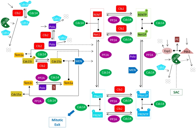
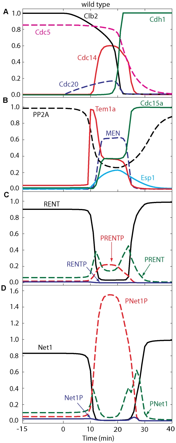
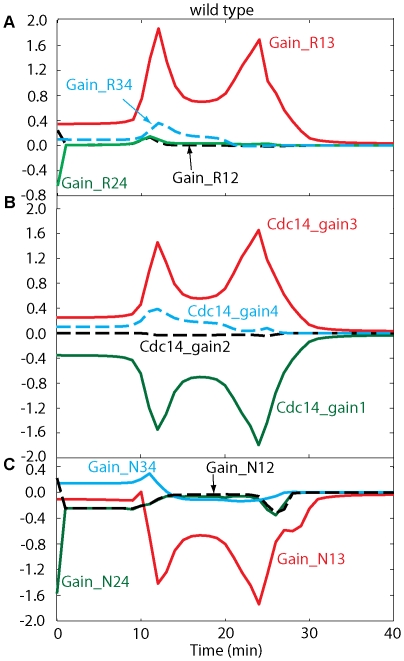
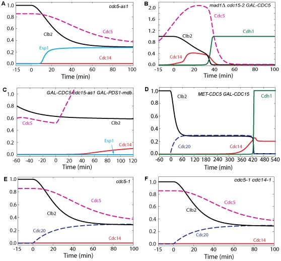
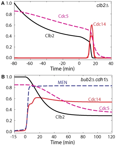
Cell cycle progression in eukaryotes is regulated by periodic activation and inactivation of a family of cyclin-dependent kinases (Cdk's). Entry into mitosis requires phosphorylation of many proteins targeted by mitotic Cdk, and exit from mitosis requires proteolysis of mitotic cyclins and dephosphorylation of their targeted proteins. Mitotic exit in budding yeast is known to involve the interplay of mitotic kinases (Cdk and Polo kinases) and phosphatases (Cdc55/PP2A and Cdc14), as well as the action of the anaphase promoting complex (APC) in degrading specific proteins in anaphase and telophase. To understand the intricacies of this mechanism, we propose a mathematical model for the molecular events during mitotic exit in budding yeast. The model captures the dynamics of this network in wild-type yeast cells and 110 mutant strains. The model clarifies the roles of Polo-like kinase (Cdc5) in the Cdc14 early anaphase release pathway and in the G-protein regulated mitotic exit network.


Conflict of interest statement

Competing Interests: The authors have declared that no competing interests exist.

Figures

Figure 1
Figure 1. Proposed wiring diagram of mitotic exit control in the budding yeast cell cycle.
For a full justification of this diagram with references, see Texts S1 and S2. Cdc28, the kinase partner of Clb2, is not shown explicitly in this diagram. Cdc20 and Cdh1 work in collaboration with the APC, which is also not shown explicitly in the diagram. All proteins (ovals) are assumed to be produced and degraded at specific rates. Four white circles represent degraded proteins. Solid lines correspond to chemical reactions, while dashed lines denote regulatory effects (enzyme catalysis). A protein sitting on a reaction arrow also represents an enzyme that catalyses the reaction. Cdc20 initiates the transition from metaphase to anaphase. Cdc14 released from PRENT and PRENTP induces exit from mitosis, i.e., activation of Cdh1 and establishment of the cell in G1 phase.
Figure 2
Figure 2. Numerical simulation of exit from mitosis in wild-type cells.
The four panels show the time courses of ME regulators during a typical Cdc20 ‘block and release’ experiment, which is simulated as follows: the simulation starts at t = −15 min under metaphase-block conditions (cdc20Δ GAL-CDC20 in glucose; k s,20 = 0), and then Cdc20 synthesis is induced (transfer to galactose; k s,20 = 0.015) at t = 0. During the Cdc20-block phase (t<0), free Cdc14 is low due to sequestration by Net1 in RENT, MEN is inactive, and Net1 and RENT are predominantly dephosphorylated because of high activity of PP2A. The steady state levels of Clb2, Cdc5, Net1, RENT and PP2A are close to 1 (arbitrary unit).
Figure 3
Figure 3. Flux diagrams in wild-type cells.
Initially cells are in the metaphase steady state by Cdc20 deprivation. Cdc20 activation at time zero (k s,20 = 0.015) induces mitotic progression through anaphase, telophase and G1. Flux definitions are given in Table S1.
Figure 4
Figure 4. Simulation of mitotic progression of cells containing overexpressed CDC5 and inactive cdc5 mutations.
(A) Cdc5 is necessary for ME. Cdc20 block-and-release was simulated as in Figure 2 with inactive Cdc5 (cdc5-as1; effpol = 0). Cdc14 is not released, nor is Cdh1 activated. (B) The MEN requirement for ME can be bypassed by overexpressed Cdc5. Cdc20 block-and-release was simulated as usual, with inactive Cdc15 (cdc15-2; effc15 = 0) and with Cdc5 overexpressed 30-fold (GAL-CDC5; k s,polo = 0.3). (C) Overexpressed Cdc5 is sufficient for Cdc14 release when FEAR and MEN are inactive. Simulation was started in an arrested steady state with initial conditions of Clb2 and Polo were set less than metaphase values to represent an earlier stage of the arrest by hydroxyurea (Clb2 = 0.8, Polo = 0.6, Poloi = 0.2, k s,b2 = 0.024, k s,polo = 0.006) and with inactive Cdc15 (effc15 = 0) for 15 min. Then Cdc5 and Pds1 overexpressions were induced at time zero (k s,polo = 0.3, k s,pds = 0.45, k d,pds′ = 0). (D) The Cdc5 requirement for Cdc14 release and ME can be bypassed by overexpression of a truncated version of Cdc15. Cdc20 block-and-release was pre-simulated for 60 min with no synthesis of either Cdc20 or Cdc5 (k s,polo = k s,20 = 0; setting also the initial conditions for Cdc5 active and inactive forms to zero) while the total concentration of Cdc15 was increased 20-fold and inhibition of Cdc15 by Cdk was reduced 1000-fold (k i,c15′ = 0.00009, CDC15T = 20). At t = 0, Cdc20 synthesis is induced as usual (k s,20 = 0.015). (E) Cdc14 is not released in cdc5-1 and cdc5-1 cdc14-1 cells in E and F. Therefore, Cdc14 release in the cdc14-1 mutant may be attributable solely to Net1 phosphorylation by Cdc5. Simulation in E was done similar to Figure 4A except that effpol was set to 0.1 for the small residual activity of Cdc5. (F) Simulation in F was done similar to A except that activity of Cdc14 was set to zero (effc14 = 0).
Figure 5
Figure 5. Simulations of mitotic progression of cells containing cdc15-2, NET1-6cdk, tem1-3, cdc28-as1, and GAL-CLB2dbΔ cdc5-as1 mutations.
(A) In MEN mutants such as cdc15-2, Cdc14 is transiently released and resequestered. Cdc20 block-and-release was presimulated with inactive Cdc15 (effc15 = 0). (B) In tem1-3 temperature sensitive mutant (MEN inactive), Cdc14 is transiently released and cells are arrested in telophase. Simulation was started at metaphase by Cdc20 deprivation (k s,20 = 0) for 15 min with total concentration of Tem1, initial conditions of Tem1 and MEN were set to zero. Cdc20 was activated at time zero (k s,20 = 0.015) (C) In NET1-6cdk cells ME occurs with a delay, as typical of FEAR mutants. Cdc20 block-and-release was presimulated with no Net1 phosphorylation by Cdk/Clb2 (k k,12 = k k,34 = 0). (D) Double MEN and FEAR mutations, such as NET1-6cdk cdc15-2, do not show transient release of Cdc14 and arrest in telophase. Cdc20 block-and-release was presimulated with inactive Cdc15 (effc15 = 0) and no Net1 phosphorylation by Cdk/Clb2 (k k,12 = k k,34 = 0). (E) When Cdk kinase activity is inhibited, there is no Cdc14 release. Both Cdk/Clb2 and Cdc5 phosphorylation on Net1 are diminished in cdc28-as1 mutant. Simulation was done similar to wild-type cells except that INH was set to 5 to inhibit Cdk kinase activity. (F) Our model predicts that when Cdc5 is inhibited, overexpressed Clb2 cannot induce Cdc14 release with or without active Cdc20. Simulation was started at metaphase by Cdc20 deprivation, overexpression of Clb2 and inactive Cdc5 for 15 min (k s,20 = 0, k s,b2 = 0.6, k d,b2 = k d,b2′ = effpol = 0). Cdc20 was added back at time zero (k s,20 = 0.015).
Figure 6
Figure 6. Flux diagrams and temporal changes of RENT, Net1 forms in NET1-6cdk mutants.
Simulation was done similar to Figure 5C.
Figure 7
Figure 7. Temporal changes of RENT, Net1 forms and fluxes in cdc15-2 cells blocked at telophase in mitosis.
Simulation was done similar to Figure 5A.
Figure 8
Figure 8. Model predicts that Cdc14 is responsible for its own re-sequestration after ME.
(A–C) All simulations were done similar to cdc15-2 mutant simulations in Figure 5A except that after 20 min either Cdc14 (in A, effc14 = 0) or PP2A (in B, effppa = 0) or both (in C, effc14 = effppa = 0)) were inactivated by setting their corresponding activity factors to zero.
Figure 9
Figure 9. Mitotic progression of cells containing an ESP1 mutation.
(A) In metaphase arrested cells at 23°C, overexpression of Esp1 induces Cdc14 release; however, cells do not exit from mitosis, and Cdh1 stays inactive. Cells are presimulated in metaphase arrest by Cdc20 deprivation, then at t = 0 the rate of synthesis of Esp1 is increased 60-fold (k s,esp = 0.078), with the rate of Clb2 degradation at 23°C assumed to be half its basal value (k d,b2″ = 1.5). (B) Cdc14 release is dependent upon Cdc5 in nocodazole-arrested cells; when CDC5 is deleted, overexpressed Esp1 can no longer induce Cdc14 release. Cells are presimulated in metaphase arrest by nocodazole (N = 1) with no synthesis of Cdc5 (k s,polo = 0) and no initial Cdc5 proteins. Then at t = 0 the rate of synthesis of Esp1 is increased 60-fold (k s,esp = 0.078). (C) When CLB5 is deleted, overexpressed Esp1 can induce ME. Reduction in Cdk activity by Clb5 deprivation allows for ME by increasing the phosphatase-to-kinase ratio, leading to activation of Cdh1. Simulation was done as in panel A, except that the synthesis rate of Clb2 was set to 80% of its basal value (k s,b2 = 0.024). (D) When Esp1 is inactive, Cdc14 cannot be released and the cell cannot exit from mitosis. It is assumed that separase is absent in esp1-2td mutant cells (ks,esp = 0). During the 120 min pre-simulation of Cdc20 block in metaphase, the rate of degradation of Esp1 was increased 10-fold, and the activity of Esp1 was lowered 10-fold. (effesp = 0.1, kd,esp = 0.028,). At t = 0, Cdc20 synthesis was induced, as usual.
Figure 10
Figure 10. Mitotic progression of cells containing CDC55, CLB2 and BUB2 mutations.
(A) In the CDC55 deletion strain, Cdc14 is re-sequestered with a delay. Cdc20 block-and-release was simulated as usual, with [PP2A]total = 0. (B) Cdc14 is released prematurely in bub2Δ cells. Cdc20 block-and-release was pre-simulated with the rates of Tem1 inactivation by Cdc14 and PP2A set to zero (k i,tem′ = k i,tem″ = 0). (C) In a nocodazole-arrested cell, overexpression of Clb2 induces Cdc14 release, and the cell arrests in telophase. Pre-simulation was done by setting N = 1 for 15 min. At t = 0, the rate of synthesis of Clb2 was increased 20-fold and the rates of degradation of Clb2 were set to zero (N = 1, k d,b2′ = k d,b2″ = 0, k s,b2 = 0.6). (D) Cdc14 is not released when Clb2 is overexpressed in G1 cells with Cdc5 inactive. This simulation was started from G1 initial conditions (low levels of Clb2, Cdc5 and Cdc14). At t = 0, the initial condition of Polo is set to 0.01, the synthesis rate of Clb2 is set to a large value and its degradation rate is set to zero (k d,b2′ = k d,b2″ = k s,polo = 0, k s,b2 = 0.6).
Figure 11
Figure 11. Inactive Clb2 is not required, whereas Polo inactivation is sufficient for Cdc14 re-sequestration to the nucleolus.
(A) Cells of the triple-deletion strain cdc20Δ pds1Δ cdh1Δ arrest in telophase with Cdc14 released from the nucleolus. Simulation was done by setting to zero the rate of synthesis of Pds1, the total concentration of Cdh1, and the initial conditions of Cdh1, Pds1 and PE complex (k s,pds = CDH1T = 0). (B) After 6 hours of telophase arrest, cdc20Δ pds1Δ cdh1Δ cells are subjected to Sic1 overexpression, and Cdc14 does not completely return to the nucleolus. Simulation was done as in panel A; after 6 hours INH was set to 5 to implement Cdk inhibition by Sic1. (C) In cdc20Δ pds1Δ cdh1Δ cells arrested in telophase, deprivation of Cdc5 causes return of Cdc14 to the nucleolus. Simulation was done as in panel A; after 40 min the rate of synthesis of Cdc5 was set to zero and the basal degradation rate of Cdc5 was increased 10-fold (k s,polo = 0, k d,polo = 0.1).
Figure 12
Figure 12. Effects of Cdc5 kinase and of Cdc14 and PP2A phosphatase activities on Cdc14 re-sequestration.
A stabilized version of Cdc5 (3×CDC5ΔN70) causes a delay in Cdc14 re-sequestration both in wild-type cells (panel A) and bub2Δ background (panel B). (A) Cdc20 block-and-release was pre-simulated for 180 min with no degradation of Cdc5 (k d,polo′ = 0, k s,polo = 0.011). (B) Simulation was done as in A with the rates of inactivation of Tem1 set to zero (k i,tem′ = k i,tem″ = 0). (C) Cdc14 stays released from the nucleolus in cdc14-1 cells arrested in telophase. Cdc20 block-and-release was presimulated with no Cdc14 activity (effc14 = 0). (D, E and F) Cdc20 block-and-release in wild-type cells; after 24 min (when cells start to enter G1 phase), either PP2A activity (panel D, effpa = 0) or Cdc14 activity (panel E, effc14 = 0) was inhibited. In either case, Cdc14 is re-sequestered into the nucleoulus. In panel F, when both PP2A and Cdc14 phosphatase activities are inhibited after 24 min (effpa = effc14 = 0), Cdc14 does not return to the nucleolus.
Figure 13
Figure 13. clb2Δ and bub2Δ cdh1Δ mutants.
(A) When Clb2 is inhibited Cdc14 is released with a delay. Simulation was started at metaphase Cdc20 block for 80 min with rate of synthesis of Clb2 in the model decreased to 1/3 of baseline due to residual Clb1 activity (k s,b2 = 0.1) and Cdc20 was added back at time zero. (B) In bub2Δ cdh1Δ cells, Cdc14 stays released after ME. Simulation was done as wild-type cells except that rates of inactivation of Tem1 and total concentration of Cdh1 were set to zero (k i,tem′ = k i,tem″ = CDH1T = 0).

Similar articles

Cited by

References

    1. Nurse P. Universal control mechanism regulating onset of M-phase. Nature. 1990;344:503–508. - PubMed
    1. Nowak MA, Komarova NL, Sengupta A, Jallepalli PV, Shih Ie M, et al. The role of chromosomal instability in tumor initiation. Proc Natl Acad Sci U S A. 2002;99:16226–16231. - PMC - PubMed
    1. Novak B, Tyson JJ. Quantitative-Analysis of a Molecular-Model of Mitotic Control in Fission Yeast. Journal of Theoretical Biology. 1995;173:283–305.
    1. Novak B, Tyson JJ. Modeling the control of DNA replication in fission yeast. Proc Natl Acad Sci U S A. 1997;94:9147–9152. - PMC - PubMed
    1. Sveiczer A, Csikasz-Nagy A, Gyorffy B, Tyson JJ, Novak B. Modeling the fission yeast cell cycle: quantized cycle times in wee1- cdc25Delta mutant cells. Proc Natl Acad Sci U S A. 2000;97:7865–7870. - PMC - PubMed

Publication types

MeSH terms

Substances

LinkOut - more resources