Disparate impact of butyroyloxymethyl diethylphosphate (AN-7), a histone deacetylase inhibitor, and doxorubicin in mice bearing a mammary tumor
- PMID: 22384017
- PMCID: PMC3285631
- DOI: 10.1371/journal.pone.0031393
Disparate impact of butyroyloxymethyl diethylphosphate (AN-7), a histone deacetylase inhibitor, and doxorubicin in mice bearing a mammary tumor
Abstract
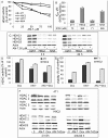
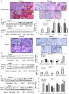
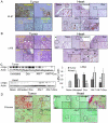
The histone deacetylase inhibitor (HDACI) butyroyloxymethyl diethylphosphate (AN-7) synergizes the cytotoxic effect of doxorubicin (Dox) and anti-HER2 on mammary carcinoma cells while protecting normal cells against their insults. This study investigated the concomitant changes occurring in heart tissue and tumors of mice bearing a subcutaneous 4T1 mammary tumor following treatment with AN-7, Dox, or their combination. Dox or AN-7 alone led to inhibition of both tumor growth and lung metastases, whereas their combination significantly increased their anticancer efficacy and attenuated Dox- toxicity. Molecular analysis revealed that treatment with Dox, AN-7, and to a greater degree, AN-7 together with Dox increased tumor levels of γH2AX, the marker for DNA double-strand breaks and decreased the expression of Rad51, a protein needed for DNA repair. These events culminated in increased apoptosis, manifested by the appearance of cytochrome-c in the cytosol. In the myocardium, Dox-induced cardiomyopathy was associated with an increase in γH2AX expression and a reduction in Rad51 and MRE11 expression and increased apoptosis. The addition of AN-7 to the Dox treatment protected the heart from Dox insults as was manifested by a decrease in γH2AX levels, an increase in Rad51 and MRE11 expression, and a diminution of cytochrome-c release. Tumor fibrosis was high in untreated mice but diminished in Dox- and AN-7-treated mice and was almost abrogated in AN-7+Dox-treated mice. By contrast, in the myocardium, Dox alone induced a dramatic increase in fibrosis, and AN7+Dox attenuated it. The high expression levels of c-Kit, Ki-67, c-Myc, lo-FGF, and VEGF in 4T1 tumors were significantly reduced by Dox or AN-7 and further attenuated by AN-7+Dox. In the myocardium, Dox suppressed these markers, whereas AN-7+Dox restored their expression. In conclusion, the combination of AN-7 and Dox results in two beneficial effects, improved anticancer efficacy and cardioprotection.
Conflict of interest statement
Figures


Similar articles
-
The histone deacetylase inhibitor butyroyloxymethyl diethylphosphate (AN-7) protects normal cells against toxicity of anticancer agents while augmenting their anticancer activity.Invest New Drugs. 2012 Feb;30(1):130-43. doi: 10.1007/s10637-010-9542-z. Epub 2010 Sep 23. Invest New Drugs. 2012. PMID: 20862515
-
AN-7, a butyric acid prodrug, sensitizes cutaneous T-cell lymphoma cell lines to doxorubicin via inhibition of DNA double strand breaks repair.Invest New Drugs. 2018 Feb;36(1):1-9. doi: 10.1007/s10637-017-0500-x. Epub 2017 Sep 8. Invest New Drugs. 2018. PMID: 28884410
-
Cardioprotection by AN-7, a prodrug of the histone deacetylase inhibitor butyric acid: Selective activity in hypoxic cardiomyocytes and cardiofibroblasts.Eur J Pharmacol. 2020 Sep 5;882:173255. doi: 10.1016/j.ejphar.2020.173255. Epub 2020 Jun 15. Eur J Pharmacol. 2020. PMID: 32553737
-
Histone deacetylase inhibitors: the anticancer, antimetastatic and antiangiogenic activities of AN-7 are superior to those of the clinically tested AN-9 (Pivanex).Clin Exp Metastasis. 2008;25(7):703-16. doi: 10.1007/s10585-008-9179-x. Epub 2008 May 28. Clin Exp Metastasis. 2008. PMID: 18506586
-
A novel valproic acid prodrug as an anticancer agent that enhances doxorubicin anticancer activity and protects normal cells against its toxicity in vitro and in vivo.Biochem Pharmacol. 2014 Mar 15;88(2):158-68. doi: 10.1016/j.bcp.2014.01.023. Epub 2014 Jan 24. Biochem Pharmacol. 2014. PMID: 24463168
Cited by
-
Qishen Yiqi Dropping Pills Ameliorates Doxorubicin-induced Cardiotoxicity in Mice via Enhancement of Cardiac Angiogenesis.Med Sci Monit. 2019 Apr 3;25:2435-2444. doi: 10.12659/MSM.915194. Med Sci Monit. 2019. PMID: 30943187 Free PMC article.
-
miR-320a mediates doxorubicin-induced cardiotoxicity by targeting VEGF signal pathway.Aging (Albany NY). 2016 Jan;8(1):192-207. doi: 10.18632/aging.100876. Aging (Albany NY). 2016. PMID: 26837315 Free PMC article.
-
The Histone Deacetylase Inhibitor AN7, Attenuates Choroidal Neovascularization in a Mouse Model.Int J Mol Sci. 2019 Feb 7;20(3):714. doi: 10.3390/ijms20030714. Int J Mol Sci. 2019. PMID: 30736437 Free PMC article.
-
The Therapeutic Potential of AN-7, a Novel Histone Deacetylase Inhibitor, for Treatment of Mycosis Fungoides/Sezary Syndrome Alone or with Doxorubicin.PLoS One. 2016 Jan 11;11(1):e0146115. doi: 10.1371/journal.pone.0146115. eCollection 2016. PLoS One. 2016. PMID: 26752418 Free PMC article.
-
Effects of histone deacetylase inhibitory prodrugs on epigenetic changes and DNA damage response in tumor and heart of glioblastoma xenograft.Invest New Drugs. 2017 Aug;35(4):412-426. doi: 10.1007/s10637-017-0448-x. Epub 2017 Mar 17. Invest New Drugs. 2017. PMID: 28315153
References
-
- Xu WS, Parmigiani RB, Marks PA. Histone deacetylase inhibitors: molecular mechanisms of action. Oncogene. 2007;26:5541–5552. - PubMed
-
- Rosato RR, Grant S. Histone deacetylase inhibitors in clinical development. Expert Opin Investig Drugs. 2004;13:21–38. - PubMed
-
- Colussi C, Illi B, Rosati J, Spallotta F, Farsetti A, et al. Histone deacetylase inhibitors: Keeping momentum for neuromuscular and cardiovascular diseases treatment. Pharmacol Res. 2010;62:3–10. - PubMed
-
- Rephaeli A, Rabizadeh E, Aviram A, Shaklai M, Ruse M, et al. Derivatives of butyric acid as potential anti-neoplastic agents. Int J Cancer. 1991;49:66–72. - PubMed
-
- Rephaeli A, Blank-Porat D, Tarasenko N, Entin-Meer M, Levovich I, et al. In vivo and in vitro antitumor activity of butyroyloxymethyl-diethyl phosphate (AN-7), a histone deacetylase inhibitor, in humoan prostate cancer. Int J Cancer. 2005;116:226–235. - PubMed
Publication types
MeSH terms
Substances
LinkOut - more resources
Full Text Sources
Other Literature Sources
Research Materials
Miscellaneous

