. 2012;7(2):e31430.
doi: 10.1371/journal.pone.0031430. Epub 2012 Feb 24.

In vitro study of novel collagenase (XIAFLEX®) on Dupuytren's disease fibroblasts displays unique drug related properties

Affiliations

In vitro study of novel collagenase (XIAFLEX®) on Dupuytren's disease fibroblasts displays unique drug related properties

Farhatullah Syed et al. PLoS One. 2012.

Abstract

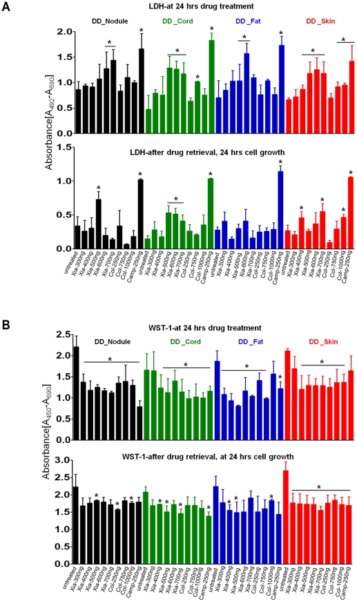
Dupuytren's disease (DD) is a benign, fibroproliferative disease of the palmar fascia, with excessive extracellular matrix (ECM) deposition and over-production of cytokines and growth factors, resulting in digital fixed flexion contractures limiting hand function and patient quality of life. Surgical fasciectomy is the gold standard treatment but is invasive and has associated morbidity without limiting disease recurrence. Injectable Collagenase Clostridium histolyticum (CCH)--Xiaflex®--is a novel, nonsurgical option with clinically proven in vivo reduction of DD contractures but with limited in vitro data demonstrating its cellular and molecular effects. The aim of this study was to delineate the effects of CCH on primary fibroblasts isolated from DD and non-DD anatomical sites (using RTCA, LDH, WST-1, FACS, qRT-PCR, ELISA and In-Cell Quantitative Western Blotting) to compare the efficacy of varying concentrations of Xiaflex® against a reagent grade Collagenase, Collagenase A. Results demonstrated that DD nodule and cord fibroblasts had greater proliferation than those from fat and skin. Xiaflex® exposure resulted in dose- and time-dependent inhibition of cellular spreading, attachment and proliferation, with cellular recovery after enzyme removal. Unlike Collagenase A, Xiaflex® did not cause apoptosis. Collagen expression patterns were significantly (p<0.05) different in DD fibroblasts across anatomical sites - the highest levels of collagen I and III were detected in DD nodule, with DD cord and fat fibroblasts demonstrating a smaller increase in both collagen expression relative to DD skin. Xiaflex® significantly (p<0.05) down-regulated ECM components, cytokines and growth factors in a dose-dependent manner. An in vitro scratch wound assay model demonstrated that, at low concentrations, Xiaflex® enabled a faster fibroblast reparatory migration into the wound, whereas, at high concentrations, this process was significantly (p<0.05) inhibited. This is the first report elucidating potential mechanisms of action of Xiaflex® on Dupuytren fibroblasts, offering a greater insight and a better understanding of its effect in DD.


Conflict of interest statement

Competing Interests: The authors have the following competing interest: Auxilium Pharmaceuticals donated Xiaflex®, SH was in the employment of Auxilium Pharmaceuticals at the time the project was undertaken. There are no patents, products in development or marketed products to declare. This does not alter the authors' adherence to all the PLoS ONE policies on sharing data and materials, as detailed online in the guide for authors.

Figures

Figure 1
Figure 1. Flowchart demonstrating the tissue collection, processing and experimental strategy used in this study.
Figure 2
Figure 2. Real-Time Monitoring of Xiaflex® and Collagenase A effect on DD primary fibroblasts from different anatomical sites using MESA.
Primary fibroblasts of DD (Nodule, Cord, Fat and Skin) were seeded onto the E-plate and cells were allowed to grow prior to the introduction of Xiaflex® and Collagenase A at various concentrations. After drug addition, cells were allowed to grow for 24 hours in the presence of drugs. After 24 hrs, the drugs were removed and the cells were fed with fresh supWillE media for 24 hrs to assess the reversibility of the inhibitory effect of the drugs. Cell Indexes were recorded every 15 minutes. Each trace at each concentration was an average of three replicates. A. Effect of Xiaflex® and Collagenase A on DD-Nodule. B. Effect of Xiaflex® and Collagenase A on DD-Cord.
Figure 3
Figure 3. Real-Time Cell Analaysis (RTCA) monitoring of Xiaflex® and Collagenase A effects on DD-primary fibroblasts obtained from different anatomical sites.
This diagram demonstrates average cell indeces (CI) of untreated and treated cell groups taken from six independent RTCA experiments, which have been plotted.
Figure 4
Figure 4. Effect of Xiaflex® and Collagenase A on cell membrane integrity (cytotoxicity detection) and cell viability/metabolic activity measured by LDH and WST-1 assays.
A. LDH (lactose dehydrogenase) leakage assay for cell membrane integrity assessed the cytotoxic effect of the drugs. B. WST-1 (water soluble-tetrazolium salt-1) assayed for cell viability/metabolic activity and cell death. *p<0.05, indicates significant difference compared to untreated group. The data was expressed as average means ± SEM from four independent experiments.
Figure 5
Figure 5. Detection of Early Apoptosis and Necrosis using Annexin V and PI.
Fibroblasts from different anatomical sites (Nodule, Cord, Fat and Skin) were treated with various concentration of Xiaflex® and Collagenase A as indicated in the graphs. 24 hours post-treatment, cells were harvested and labeled with Annexin V-FITC and PI. A. FITC-conjugated annexin V staining for untreated cells, upper left plot (labeled-untreated), compared with the viable control cells, upper right plot (unlabeled cells). Dual-staining of treated cells (lower panel): the quadrant analysis shows viable cells negative for annexin V and PI in the lower left, R3. Apoptotic cells stained with annexin V but excluding PI are shown in the lower right, R4. Secondary necrotic cells (i.e. necrosis after apoptosis) positive for both PI and annexin V are shown in upper right, R2. Necrotic or mechanically damaged cells positive for PI only are shown in upper left, R1. Representative data are shown from three independent experiments in triplicates. B. Annexin V and PI positive cells after 24 hrs treatment with Xiaflex® and Collagenase A at various concentrations as indicated in bar graph. Positive cells were counted from three independent experiments and plotted on the graph as an average means ± SEM. *p<0.05, indicates significant difference compared to untreated group.
Figure 6
Figure 6. Measurement of comparative effect of Xiaflex and Collagenase A on mRNA steady-state levels of primary DD fibroblasts isolated from different anatomical sites.
A. Collagen I; B. Collagen III. #p<0.05 indicates significant difference in the expression of collagen I & III in the Nodule compared to the Cord, Fat and Skin fibroblasts. +p<0.05 indicates significant difference in the expression of collagen I in Cord compared to Fat and Skin fibroblasts. ++p<0.05 indicates significant difference in the expression of collagen I in Fat compared to Skin fibroblasts. &p<0.05 indicates significant difference in the expression of collagen III in Cord compared to Skin fibroblasts. $p<0.05 indicates significant difference in the expression of collagen III in Fat compared to Cord and Skin fibroblasts. C. Fibronectin; D. α-SMA; E. TGF-β I; F. MMP-2; G. MMP-9. *p<0.05, **p≤0.01 indicates significant difference between mRNA steady state levels of untreated and treated fibroblasts. The data presented here are the averaged results from three independent experiments.
Figure 7
Figure 7. In vitro quantitative measurement of collagen I and III in DD fibroblasts.
Synthesis of collagen I and III was measured by capture sandwich ELISA (for collagen I expression) and Indirect ELISA (for collagen III expression). *p<0.05, **p≤0.01 indicates significant difference compared to the DD-Skin fibroblasts. #p<0.05 indicates significant difference in Nodule compared to the Cord, Fat and Skin fibroblasts, in the expression of collagen I. ##p≤0.01 indicates significant difference in the expression of collagen III in Nodule compared to Cord, Fat and Skin fibroblasts. The data presented here are the means ± SEM of three averaged independent experiments carried out in triplicates.
Figure 8
Figure 8. Comparison of the effect of Xiaflex® and Collagenase A on Type I and Type III collagen protein synthesis by DD fibroblasts.
DD fibroblasts from passages 1–4 were cultured in a 96 well plate (1.5×10⁴cells/well) and treated with both drugs at various concentrations as indicated in the figure. Cell lysates from treated and untreated cells were subjected to capture sandwich ELISA (for the detection of collagen I) and Indirect ELISA (for the detection of collagen III) as described previously. A. Collagen I and B. Collagen III. *p<0.05, **p≤0.01, indicate significant difference in treated group compared to untreated control group. The data presented here are the average of three independent experiments performed in triplicates.
Figure 9
Figure 9. Quantitative In-Cell Western blotting assay for the analysis of protein expression upon treatment with Xiaflex® and Collagenase A by DD fibroblasts.
Primary fibroblasts were seeded in 96 well plate (1.5×10⁴cells/well) and allowed to grow for ∼7–8 hours. The cells were then treated with various concentrations of Xiaflex® and Collagenase A as indicated. At 24 hours drug treatment, the cells were fixed in 4% formaldehyde/PBS. In-Cell Western blotting was performed. Representative output infrared images of treated and untreated fibroblasts, stained for protein expression (visible in green/red) from different anatomical sites are shown. Bar graphs represent the quantification of average protein expression in different treatments from three independent experiments after normalization to loading control beta-actin. A. Collagen I; B. Collagen III; C. Fibronectin; D. α-SMA; E. Desmin; F. Tenascin; G. Collagen IV; H. CTGF; I. MMP-9. *p<0.05, **p≤0.01, indicate significant difference in treated group compared to untreated control group. The data presented here are the means ± SEM of triplicate experiments.unt: untreated.
Figure 10
Figure 10. Comparison of the effect of Xiaflex® and Collagenase A on cell cycle regulation.
Cell cycle gene (PCNA, Cyclin D1 and Cyclin D2) were assessed at mRNA and protein levels using qRT-PCR and In-Cell Western blotting respectively. A. mRNA steady-state levels of cell cycle genes (PCNA, Cyclin D1 and Cyclin D2) after treatment with Xiaflex® and Collagenase A at various concentrations as indicated in the graphs. All the cell cycle genes were dose-dependently down regulated by Xiaflex® and Collagenase A compared to the untreated control group. B. Relative protein expression of cell cycle proteins (PCNA and Cyclin D) after treatment with Xiaflex® and Collagenase A. *p<0.05, **p≤0.01, indicate significant difference in treated group compared to untreated control group. The data presented here are the average of three independent experiments performed in triplicate. Results are presented as means ± SEM of triplicates.
Figure 11
Figure 11. Effect of Xiaflex® on DD fibroblast migration towards a mechanically created in vitro scratch wound.
A. Fibroblast migration response towards injury with and without drugs. Representative micrographs are shown from three independent experiments. B. Number of migrated fibroblasts towards wound with and without drugs. Migrated cells into the wound area were counted based on the 0 hour migration pattern from four micrographs for each treatment. Average of number of migrated cells, from three independent experiments carried out in triplicate were plotted on the graph. *p<0.05, **p≤0.01, indicate significant difference in the treated group compared to untreated control (WE+no_Drug) group. #p<0.05 indicates significant difference compared to all the groups.

Similar articles

Cited by

References

    1. Bayat A, McGrouther DA. Management of Dupuytren's disease - Clear advice for an elusive condition. Annals of the Royal College of Surgeons of England. 2006;88:3–8. - PMC - PubMed
    1. Thurston AJ. Dupuytren's disease. Journal of Bone and Joint Surgery - Series B. 2003;85:469–477. - PubMed
    1. Caufield RJ, Edwards SG. Dupuytren disease: An update on recent literature. Current Orthopaedic Practice. 2008;19:499–502.
    1. Dupuytren G. Permanent retraction of the fingers, produced by an affection of the palmar fascia. Lancet. 1834;2:222–225.
    1. Shih B, Bayat A. Scientific understanding and clinical management of Dupuytren disease. Nat Rev Rheumatol. 2010;6:715–726. - PubMed

Publication types

MeSH terms

Substances