

Comparison of functional proteomic analyses of human breast cancer cell lines T47D and MCF7
- PMID: 22384035
- PMCID: PMC3286449
- DOI: 10.1371/journal.pone.0031532
Comparison of functional proteomic analyses of human breast cancer cell lines T47D and MCF7
Erratum in
- PLoS One. 2012;7(4): doi/10.1371/annotation/18f08a33-35e1-4bf9-8d21-476757dccbef. Adjo Aka, Juliette [corrected to Aka, Juliette Adjo].
Abstract
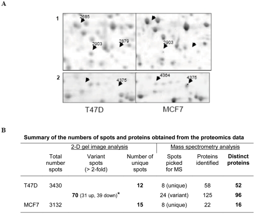
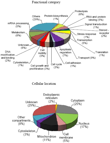
T47D and MCF7 are two human hormone-dependent breast cancer cell lines which are widely used as experimental models for in vitro and in vivo (tumor xenografts) breast cancer studies. Several proteins involved in cancer development were identified in these cell lines by proteomic analyses. Although these studies reported the proteomic profiles of each cell line, until now, their differential protein expression profiles have not been established. Here, we used two-dimensional gel and mass spectrometry analyses to compare the proteomic profiles of the two cell lines, T47D and MCF7. Our data revealed that more than 164 proteins are differentially expressed between them. According to their biological functions, the results showed that proteins involved in cell growth stimulation, anti-apoptosis mechanisms and cancerogenesis are more strongly expressed in T47D than in MCF7. These proteins include G1/S-specific cyclin-D3 and prohibitin. Proteins implicated in transcription repression and apoptosis regulation, including transcriptional repressor NF-X1, nitrilase homolog 2 and interleukin-10, are, on the contrary, more strongly expressed in MCF7 as compared to T47D. Five proteins that were previously described as breast cancer biomarkers, namely cathepsin D, cathepsin B, protein S100-A14, heat shock protein beta-1 (HSP27) and proliferating cell nuclear antigen (PCNA), are found to be differentially expressed in the two cell lines. A list of differentially expressed proteins between T47D and MCF7 was generated, providing useful information for further studies of breast cancer mechanisms with these cell lines as models.
Conflict of interest statement
Figures
Similar articles
-
Comparison of proteomic and genomic analyses of the human breast cancer cell line T47D and the antiestrogen-resistant derivative T47D-r.Mol Cell Proteomics. 2004 Jan;3(1):43-55. doi: 10.1074/mcp.M300047-MCP200. Epub 2003 Oct 13. Mol Cell Proteomics. 2004. PMID: 14557597
-
17beta-hydroxysteroid dehydrogenase type 1 modulates breast cancer protein profile and impacts cell migration.Breast Cancer Res. 2012 Jun 12;14(3):R92. doi: 10.1186/bcr3207. Breast Cancer Res. 2012. PMID: 22691413 Free PMC article.
-
Identification of 14-3-3sigma as a contributor to drug resistance in human breast cancer cells using functional proteomic analysis.Cancer Res. 2006 Mar 15;66(6):3248-55. doi: 10.1158/0008-5472.CAN-05-3801. Cancer Res. 2006. PMID: 16540677
-
Breast cancer biomarkers: proteomic discovery and translation to clinically relevant assays.Expert Rev Proteomics. 2012 Dec;9(6):599-614. doi: 10.1586/epr.12.62. Expert Rev Proteomics. 2012. PMID: 23256671 Review.
-
Breast tumor metastasis: analysis via proteomic profiling.Expert Rev Proteomics. 2008 Jun;5(3):457-67. doi: 10.1586/14789450.5.3.457. Expert Rev Proteomics. 2008. PMID: 18532913 Free PMC article. Review.
Cited by
-
Nutraceuticals known to promote hair growth do not interfere with the inhibitory action of tamoxifen in MCF7, T47D and BT483 breast cancer cell lines.PLoS One. 2024 Feb 26;19(2):e0297080. doi: 10.1371/journal.pone.0297080. eCollection 2024. PLoS One. 2024. PMID: 38408073 Free PMC article.
-
Quantitative CK19 biomarker detection in breast cancer cell lines.J Med Life. 2022 Feb;15(2):188-195. doi: 10.25122/jml-2021-1101. J Med Life. 2022. PMID: 35419102 Free PMC article.
-
Co-Adjuvant Therapy Efficacy of Catechin and Procyanidin B2 with Docetaxel on Hormone-Related Cancers In Vitro.Int J Mol Sci. 2021 Jul 2;22(13):7178. doi: 10.3390/ijms22137178. Int J Mol Sci. 2021. PMID: 34281228 Free PMC article.
-
A reappraisal of cell cycle phase enrichment in synchronized estrogen receptor-positive cell models derived from breast adenocarcinomas.Sci Rep. 2025 Feb 18;15(1):5949. doi: 10.1038/s41598-025-90456-8. Sci Rep. 2025. PMID: 39966575 Free PMC article.
-
Bioguided Fractionation of Local Plants against Matrix Metalloproteinase9 and Its Cytotoxicity against Breast Cancer Cell Models: In Silico and In Vitro Study (Part II).Molecules. 2021 Mar 8;26(5):1464. doi: 10.3390/molecules26051464. Molecules. 2021. PMID: 33800366 Free PMC article.
References
-
- Sasco AJ. Breast Cancer and the Environment. Horm Res. 2003;60(Suppl 3):50. - PubMed
-
- ESHRE Capri Workshop Group. Hormones and breast cancer. Hum Reprod Update. 2004;10:281–293. - PubMed
-
- Jemal A, Siegel R, Ward E, Hao Y, Xu J, et al. Cancer statistics, 2008. CA Cancer J Clin. 2008;58:71–96. - PubMed
-
- Canadian Cancer Society's Steering Committee. Canadian Cancer Statistics 2009. Toronto, Canada, Canadian Cancer Society 2009
-
- Husen B, Huhtinen K, Saloniemi T, Messinger J, Thole, et al. Human hydroxysteroid (17-beta) dehydrogenase 1 expression enhances estrogen sensitivity of MCF-7 breast cancer cell xenografts. Endocrinology. 2006;147:5333–5339. - PubMed
Publication types
MeSH terms
Substances
Grants and funding
LinkOut - more resources
Full Text Sources
Medical
Research Materials
Miscellaneous
