

IKs protects from ventricular arrhythmia during cardiac ischemia and reperfusion in rabbits by preserving the repolarization reserve
- PMID: 22384037
- PMCID: PMC3285162
- DOI: 10.1371/journal.pone.0031545
IKs protects from ventricular arrhythmia during cardiac ischemia and reperfusion in rabbits by preserving the repolarization reserve
Abstract
Introduction: The function of the repolarization reserve in the prevention of ventricular arrhythmias during cardiac ischemia/reperfusion and the impact of ischemia on slowly activated delayed rectifier potassium current (I(Ks)) channel subunit expression are not well understood.
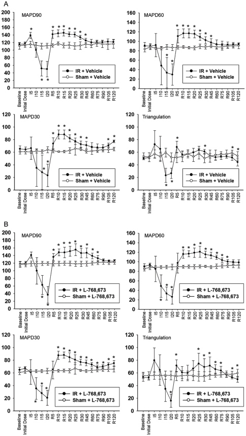
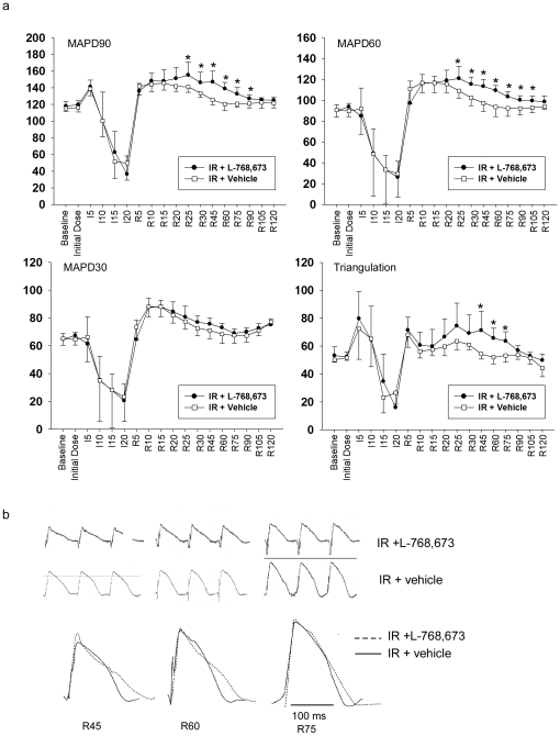
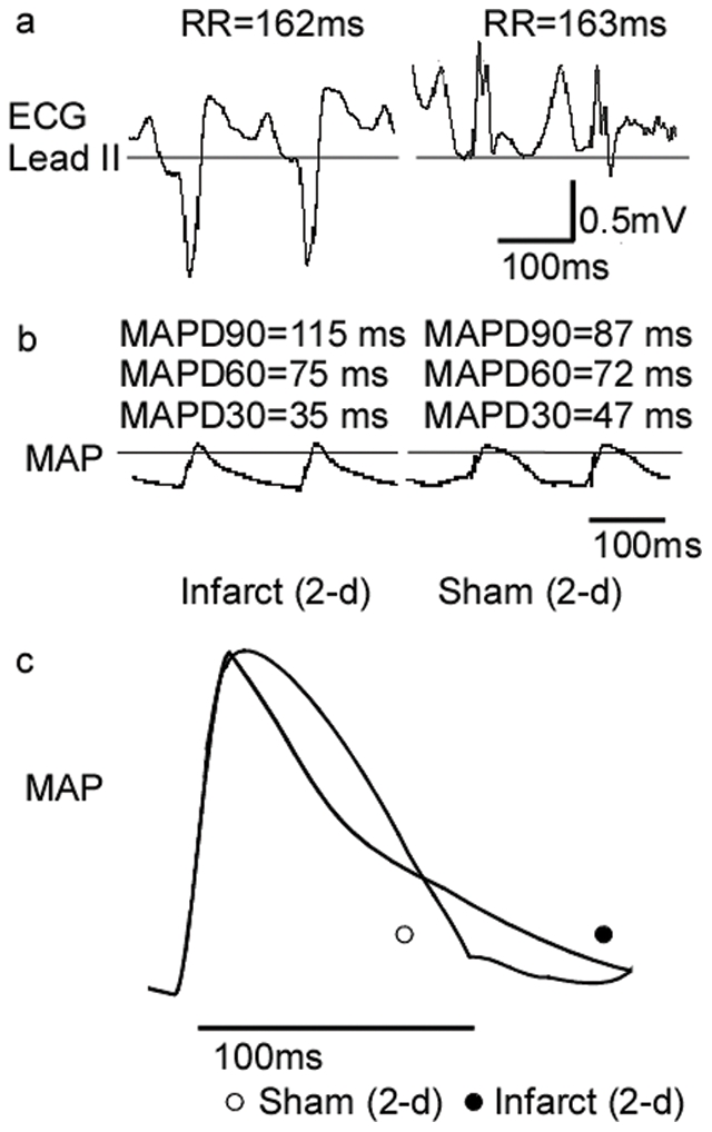
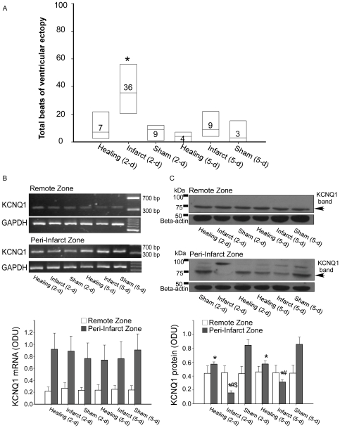
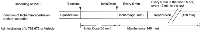
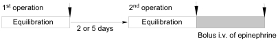
Methods and results: The responses of monophasic action potential duration (MAPD) prolongation and triangulation were investigated following an L-768,673-induced blockade of I(Ks) with or without ischemia/reperfusion in a rabbit model of left circumflex coronary artery occlusion/reperfusion. Ischemia/reperfusion and I(Ks) blockade were found to significantly induce MAPD90 prolongation and increase triangulation at the epicardial zone at 45 min, 60 min, and 75 min after reperfusion, accompanied with an increase in premature ventricular beats (PVBs) during the same period. Additionally, I(Ks) channel subunit expression was examined following transient ischemia or permanent infarction and changes in monophasic action potential (MAP) waveforms challenged by β-adrenergic stimulation were evaluated using a rabbit model of transient or chronic cardiac ischemia. The epicardial MAP in the peri-infarct zone of hearts subjected to infarction for 2 days exhibited increased triangulation under adrenergic stimulation. KCNQ1 protein, the α subunit of the I(Ks) channel, was downregulated in the same group. Both findings were consistent with an increased incidence of PVBs.
Conclusion: Blockade of I(Ks) caused MAP triangulation, which precipitated ventricular arrhythmias. Chronic ischemia increased the incidence of ventricular arrhythmias under adrenergic stimulation and was associated with increased MAP triangulation of the peri-infarct zone. Downregulation of KCNQ1 protein may be the underlying cause of these changes.
Conflict of interest statement
Figures
Similar articles
-
Paroxysmal beta-adrenergic receptor-mediated alterations in ventricular repolarization at rapid heart rates during inhibition of delayed rectifier currents.J Cardiovasc Pharmacol. 2009 Sep;54(3):253-62. doi: 10.1097/FJC.0b013e3181b2b706. J Cardiovasc Pharmacol. 2009. PMID: 19620881
-
Modulation of KCNQ1 alternative splicing regulates cardiac IKs and action potential repolarization.Heart Rhythm. 2013 Aug;10(8):1220-8. doi: 10.1016/j.hrthm.2013.04.014. Epub 2013 Apr 19. Heart Rhythm. 2013. PMID: 23608591 Free PMC article.
-
Antiarrhythmic efficacy of combined I(Ks) and beta-adrenergic receptor blockade.J Pharmacol Exp Ther. 2002 Jul;302(1):283-9. doi: 10.1124/jpet.302.1.283. J Pharmacol Exp Ther. 2002. PMID: 12065728
-
Two components of delayed rectifier K+ current in heart: molecular basis, functional diversity, and contribution to repolarization.Acta Pharmacol Sin. 2004 Feb;25(2):137-45. Acta Pharmacol Sin. 2004. PMID: 14769199 Review.
-
Pharmacological modulation of I(Ks): potential for antiarrhythmic therapy.Curr Med Chem. 2004 Jan;11(1):29-44. doi: 10.2174/0929867043456214. Curr Med Chem. 2004. PMID: 14754424 Review.
Cited by
-
In silico study of the mechanisms of hypoxia and contractile dysfunction during ischemia and reperfusion of hiPSC cardiomyocytes.Dis Model Mech. 2024 Apr 1;17(4):dmm050365. doi: 10.1242/dmm.050365. Epub 2024 Apr 26. Dis Model Mech. 2024. PMID: 38516812 Free PMC article.
-
Mechanisms Underlying Interactions Between Low-Frequency Oscillations and Beat-to-Beat Variability of Celullar Ventricular Repolarization in Response to Sympathetic Stimulation: Implications for Arrhythmogenesis.Front Physiol. 2019 Aug 2;10:916. doi: 10.3389/fphys.2019.00916. eCollection 2019. Front Physiol. 2019. PMID: 31427979 Free PMC article.
-
Insights into Cardiac IKs (KCNQ1/KCNE1) Channels Regulation.Int J Mol Sci. 2020 Dec 11;21(24):9440. doi: 10.3390/ijms21249440. Int J Mol Sci. 2020. PMID: 33322401 Free PMC article. Review.
-
Slowly activating voltage-gated potassium current potentiation by ML277 is a novel cardioprotective intervention.PNAS Nexus. 2023 May 10;2(5):pgad156. doi: 10.1093/pnasnexus/pgad156. eCollection 2023 May. PNAS Nexus. 2023. PMID: 37234204 Free PMC article.
-
Transcripts of Kv7.1 and MinK channels and slow delayed rectifier K+ current (IKs) are expressed in zebrafish (Danio rerio) heart.Pflugers Arch. 2018 Dec;470(12):1753-1764. doi: 10.1007/s00424-018-2193-1. Epub 2018 Aug 16. Pflugers Arch. 2018. PMID: 30116893
References
-
- Roden DM, Yang T. Protecting the heart against arrhythmias: potassium current physiology and repolarization reserve. Circulation. 2005;112:1376–1378. - PubMed
-
- Volders PG, Stengl M, van Opstal JM, Gerlach U, Spatjens RL, et al. Probing the contribution of IKs to canine ventricular repolarization: key role for beta-adrenergic receptor stimulation. Circulation. 2003;107:2753–2760. - PubMed
-
- Jost N, Virag L, Bitay M, Takacs J, Lengyel C, et al. Restricting excessive cardiac action potential and QT prolongation: a vital role for IKs in human ventricular muscle. Circulation. 2005;112:1392–1399. - PubMed
-
- Carmeliet E. Cardiac ionic currents and acute ischemia: from channels to arrhythmias. Physiol Rev. 1999;79:917–1017. - PubMed
Publication types
MeSH terms
Substances
LinkOut - more resources
Full Text Sources
Medical
