. 2012;7(2):e31600.
doi: 10.1371/journal.pone.0031600. Epub 2012 Feb 27.

Interleukin-1 stimulates ADAM17 through a mechanism independent of its cytoplasmic domain or phosphorylation at threonine 735

Affiliations

Interleukin-1 stimulates ADAM17 through a mechanism independent of its cytoplasmic domain or phosphorylation at threonine 735

Katherine C Hall et al. PLoS One. 2012.

Abstract

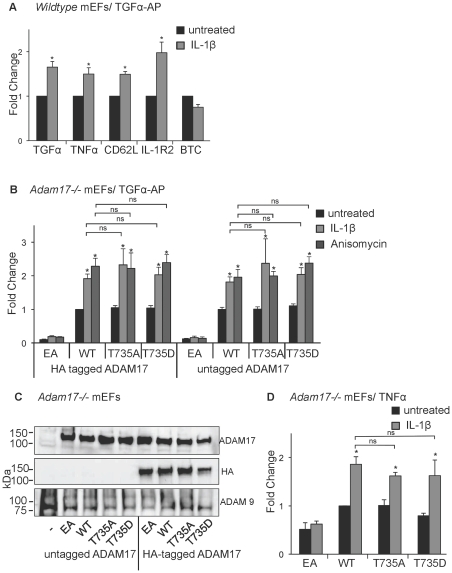
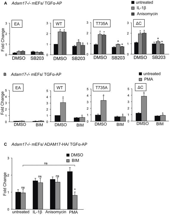
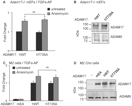
ADAM17 (a disintegrin and metalloproteinase) is a membrane-anchored metalloproteinase that regulates the release of EGFR-ligands, TNFα and other membrane proteins from cells. ADAM17 can be rapidly activated by a variety of signaling pathways, yet little is known about the underlying mechanism. Several studies have demonstrated that the cytoplasmic domain of ADAM17 is not required for its rapid activation by a variety of stimuli, including phorbol esters, tyrosine kinases and some G-protein coupled receptors. However, phosphorylation of cytoplasmic residue T735 was recently reported as a crucial step for activation of ADAM17 by IL-1β and by the p38 MAP-kinase pathway. One possible mechanism to reconcile these results would be that T735 has an inhibitory role and that it must be phosphorylated as a pre-requisite for the activation of ADAM17, which would then proceed via a mechanism that is independent of its cytoplasmic domain. To test this hypothesis, we performed rescue experiments of Adam17-/- cells with wild type and mutant forms of ADAM17. However, these experiments showed that an inactivating mutation (T735A) or an activating mutation (T735D) of cytoplasmic residue T735 or the removal of the cytoplasmic domain of ADAM17 did not significantly affect the stimulation of ADAM17 by IL-1β or by activation of MAP-kinase with anisomycin. Moreover, we found that the MAP-kinase inhibitor SB203580 blocked activation of cytoplasmic tail-deficient ADAM17 and of the T735A mutant by IL-1β or by anisomycin, providing further support for a model in which the activation mechanism of ADAM17 does not rely on its cytoplasmic domain or phosphorylation of T735.


Conflict of interest statement

Competing Interests: The authors have declared that no competing interests exist.

Figures

Figure 1
Figure 1. Stimulation of wild type and T735 mutant forms of ADAM17 by IL-1ß and anisomycin.
A) Wild type mEFs were transfected with alkaline phosphatase-tagged substrates to monitor the sheddase activity of ADAM17 (TGFα, TNFα, CD62L and IL-1R2) or ADAM10 (BTC) and stimulated with IL-1ß (5 ng/ml) for 30 minutes. The release of the AP-tagged ADAM10 or ADAM17 substrates into the culture supernatant was determined as described in materials and methods. B) Adam17−/− primary mEFs were transfected with TGFα-AP and either HA-tagged or untagged forms of the catalytically inactive ADAM17-E406A (EA), wild type ADAM17 (WT), or ADAM17 carrying point mutations at residue T735 (T735A or T735D), as indicated. The amount of TGFα-AP released in 30 minutes from untreated cells (black bars) or from cells stimulated with IL-1ß (5 ng/ml, light grey bars) or anisomycin (1 µM, dark grey bars) was determined as described in materials and methods. C) Western blots of untagged or HA-tagged forms of ADAM17 (EA, WT, T735A or T735D) expressed in Adam17−/− mEFs were probed with antibodies against the cytoplasmic domain of ADAM17 (top panel) or the HA-tag (middle panel), or against ADAM9 as a loading control. D) Shedding of transfected human TNFα from Adam17−/− mEFs rescued with WT or the T735A, T735D or EA mutant forms of ADAM17. The amount of TNFα released into the conditioned media from unstimulated cells or cells stimulated with IL-1β (5 ng/ml) in 30 minutes was determined by ELISA. All data represent the average of at least three separate experiments, and the error bars correspond to +/− standard error of the mean (sem). Asterisks indicate a significant increase upon addition of the stimulatory agent (students t-test, p<0.05). NS indicates that no significant difference was found between the indicated conditions.
Figure 2
Figure 2. Effect of inhibitors of p38 MAPK or PKC on stimulated shedding by ADAM17T735A and ADAM17ΔC.
A) Adam17−/− mEFs were co-transfected with TGFα-AP and murine WT ADAM17, EA, T735A or ΔC mutants, and either not treated (black bars) or stimulated with IL-1β (5 ng/ml, light grey bars) or anisomycin (1 µM, dark grey bars), in the presence or absence of 10 µM of the p38 MAPK inhibitor SB203580 (SB203) as indicated, with DMSO serving as a control. Constitutive and stimulated shedding of TGFα-AP was rescued by cotransfection with WT ADAM17 as well as the T735A and ΔC mutants, but not by the inactive EA mutant. There was no significant difference between the constitutive or stimulated shedding of TGFα-AP from Adam17−/− mEFs rescued with WT compared to the T735A or ΔC mutant of ADAM17. IL-1β and anisomycin-stimulated shedding of TGFα-AP in cells rescued with WT, T735 or ΔC was blocked to a comparable extent by 10 µM of the p38 MAPK inhibitor SB203580. B) TGFα-AP shedding from Adam17−/− cells rescued with WT ADAM17, T735A or ΔC was stimulated to a comparable level by 25 ng/ml PMA (untreated, black bars, PMA-treated, grey bars), and the stimulation could be blocked by the PKC inhibitor BIM I (4 µM). C) Adam17−/− mEFs transfected with TGFα together with WT ADAM17 were stimulated with 5 ng/ml IL-1β, 1 µM anisomycin or 25 ng/ml PMA in the presence or absence of 4 µM BIM I. BIM I blocked the TGFα-AP shedding stimulated by PMA, but had no significant effect on the shedding stimulated by IL-1β or anisomycin. All results represent the average values of at least 3 experiments, and the error bars indicate +/− standard error of the mean (sem). Asterisks indicate a significant increase upon addition of a stimulus, whereas plus (+) indicates a significant decrease in stimulated shedding upon introduction of an inhibitor (students t-test, p<0.05).
Figure 3
Figure 3. Comparison of TGFα shedding by human wild type ADAM17 and the T735A mutant.
A) Adam17−/− mEFs were transfected with TGFα-AP together with human wild type ADAM17 (hWT) or the human ADAM17-T735A mutant (hT735A). TGFα-AP shedding was measured in supernatants conditioned for 30 minutes on untreated cells (black bars) or cells treated with 1 µM anisomycin (grey bars). There was no significant difference between constitutive or anisomycin-stimulated shedding by hT735A compared to hWT ADAM17. B) Western blot analysis with antibodies against the cytoplasmic domain of ADAM17 demonstrated comparable expression of hWT and hT735A, with ADAM9 serving as a loading control. C) M2 CHO cells carrying inactivating mutations in both alleles of ADAM17 were transfected with TGFα-AP and the inactive mouse ADAM17EA to provide a negative control, or hWT or hT735A. Basal shedding of TGFα-AP was restored to comparable levels by the introduction of either hWT or hT735A. The stimulation of hWT and hT735 by 1 µM anisomycin was weaker in M2 CHO cells than in Adam17−/− mEFs rescued with these constructs, but there was no significant difference between the anisomycin-stimulated shedding by these two constructs. D) Western blot analysis of M2 CHO cells with anti-ADAM17 cytotail antibodies shows the expression of the endogenous mutant forms of ADAM17 in untransfected cells, and similar expression levels of murine ADAM17EA (mEA) compared to human WT or T735A ADAM17. The results in panels A and C represent the average of at least 3 experiments +/− sem. Asterisks indicate significant increase upon addition of a stimulus.

Similar articles

Cited by

References

    1. Black R, Rauch CT, Kozlosky CJ, Peschon JJ, Slack JL, et al. A metalloprotease disintegrin that releases tumour-necrosis factor-α from cells. Nature. 1997;385:729–733. - PubMed
    1. Horiuchi K, Kimura T, Miyamoto T, Takaishi H, Okada Y, et al. Cutting Edge: TNF-α-Converting Enzyme (TACE/ADAM17) Inactivation in Mouse Myeloid Cells Prevents Lethality from Endotoxin Shock. J Immunol. 2007;179:2686–2689. - PubMed
    1. Moss ML, Jin S-LC, Milla ME, Burkhart W, Cartner HL, et al. Cloning of a disintegrin metalloproteinase that processes precursor tumour-recrosis factor-α. Nature. 1997;385:733–736. - PubMed
    1. Bell J, Herrera AH, Li Y, Walcheck B. Role of ADAM17 in the ectodomain shedding of TNF-α and its receptors by neutrophils and macrophages. J Leukoc Biol. 2007;82 - PubMed
    1. Blobel CP. ADAMs: key players in EGFR-signaling, development and disease. Nat Rev Mol Cell Bio. 2005;6:32–43. - PubMed

Publication types