

Exploiting magnetic resonance angiography imaging improves model estimation of BOLD signal
- PMID: 22384043
- PMCID: PMC3285158
- DOI: 10.1371/journal.pone.0031612
Exploiting magnetic resonance angiography imaging improves model estimation of BOLD signal
Abstract
The change of BOLD signal relies heavily upon the resting blood volume fraction ([Formula: see text]) associated with regional vasculature. However, existing hemodynamic data assimilation studies pretermit such concern. They simply assign the value in a physiologically plausible range to get over ill-conditioning of the assimilation problem and fail to explore actual [Formula: see text]. Such performance might lead to unreliable model estimation. In this work, we present the first exploration of the influence of [Formula: see text] on fMRI data assimilation, where actual [Formula: see text] within a given cortical area was calibrated by an MR angiography experiment and then was augmented into the assimilation scheme. We have investigated the impact of [Formula: see text] on single-region data assimilation and multi-region data assimilation (dynamic cause modeling, DCM) in a classical flashing checkerboard experiment. Results show that the employment of an assumed [Formula: see text] in fMRI data assimilation is only suitable for fMRI signal reconstruction and activation detection grounded on this signal, and not suitable for estimation of unobserved states and effective connectivity study. We thereby argue that introducing physically realistic [Formula: see text] in the assimilation process may provide more reliable estimation of physiological information, which contributes to a better understanding of the underlying hemodynamic processes. Such an effort is valuable and should be well appreciated.
Conflict of interest statement
Figures

 : Primary visual cortex;
  : Visual area
  ).
).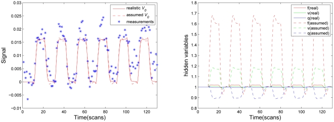
 . Real
  value is
  .
.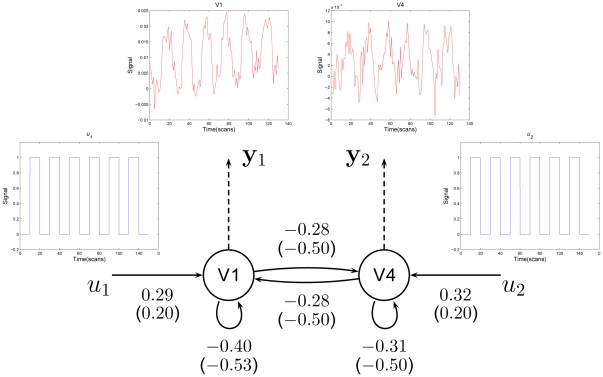
 are shown alongside the corresponding connections. The values in brackets are parameters estimated with assumed
  .
.  in visual area V
 ,
,  in V
in V and assumed
  in two areas.
  and
and  represent external inputs into the system;
  and
and  are the hemodynamic observations and arrows indicate connections.
Similar articles
-
Nonlinear estimation of BOLD signals with the aid of cerebral blood volume imaging.Biomed Eng Online. 2016 Feb 20;15:22. doi: 10.1186/s12938-016-0137-6. Biomed Eng Online. 2016. PMID: 26897355 Free PMC article.
-
Detrend-free hemodynamic data assimilation of two-stage Kalman estimator.Med Image Comput Comput Assist Interv. 2011;14(Pt 2):242-50. doi: 10.1007/978-3-642-23629-7_30. Med Image Comput Comput Assist Interv. 2011. PMID: 21995035
-
Quantitative evaluation of activation state in functional brain imaging.Brain Topogr. 2012 Oct;25(4):362-73. doi: 10.1007/s10548-012-0230-5. Epub 2012 May 9. Brain Topogr. 2012. PMID: 22569644
-
The role of susceptibility weighted imaging in functional MRI.Neuroimage. 2012 Aug 15;62(2):923-9. doi: 10.1016/j.neuroimage.2012.01.020. Epub 2012 Jan 8. Neuroimage. 2012. PMID: 22245649 Review.
-
Blood oxygenation level-dependent (BOLD)-based techniques for the quantification of brain hemodynamic and metabolic properties - theoretical models and experimental approaches.NMR Biomed. 2013 Aug;26(8):963-86. doi: 10.1002/nbm.2839. Epub 2012 Aug 28. NMR Biomed. 2013. PMID: 22927123 Free PMC article. Review.
Cited by
-
Reliable and efficient approach of BOLD signal with dual Kalman filtering.Comput Math Methods Med. 2012;2012:961967. doi: 10.1155/2012/961967. Epub 2012 Sep 10. Comput Math Methods Med. 2012. PMID: 22997541 Free PMC article.
-
An FPGA-based analysis of trade-offs in the presence of ill-conditioning and different precision levels in computations.PLoS One. 2020 Jun 19;15(6):e0234293. doi: 10.1371/journal.pone.0234293. eCollection 2020. PLoS One. 2020. PMID: 32559235 Free PMC article.
-
The Brain Effective Connectivity of Chinese during Rhyming Task.PLoS One. 2016 Sep 1;11(9):e0162158. doi: 10.1371/journal.pone.0162158. eCollection 2016. PLoS One. 2016. PMID: 27583349 Free PMC article.
-
Nonlinear estimation of BOLD signals with the aid of cerebral blood volume imaging.Biomed Eng Online. 2016 Feb 20;15:22. doi: 10.1186/s12938-016-0137-6. Biomed Eng Online. 2016. PMID: 26897355 Free PMC article.
-
Associations of resting-state fMRI functional connectivity with flow-BOLD coupling and regional vasculature.Brain Connect. 2015 Apr;5(3):137-46. doi: 10.1089/brain.2014.0299. Epub 2015 Jan 9. Brain Connect. 2015. PMID: 25384681 Free PMC article.
References
-
- Buxton RB, Wong EC, Frank LR. Dynamics of Blood Flow and Oxygenation Changes During Brain Activation: The Balloon Model. Magnetic Resonance in Medicine. 1998;39:855–864. - PubMed
-
- Buxton RB, Frank LR. A model for the coupling between cerebral blood flow and oxygen metabolism during neural stimulation. Journal of Cerebral Blood & Flow Metabolism. 1997;17:64–72. - PubMed
-
- Friston KJ, Mechelli A, Turner R, Price CJ. Nonlinear Responses in fMRI: The Balloon Model, Volterra Kernels, and Other Hemodynamics. NeuroImage. 2000;12:466–477. - PubMed
-
- Riera JJ, Watanabe J, Kazuki I, Naoki M, Aubert E, et al. A State-space Model of the Hemodynamic Approach: Nonlinear Filtering of BOLD Signals. NeuroImage. 2004;21:547–567. - PubMed
-
- Johnston LA, Duff E, Egan GF. Particle Filtering for Nonlinear BOLD Signal Analysis. 2006. pp. 292–299. In: 9th International Conference on Medical Image Computing and Computer Assisted Intervention (MICCAI). Copenhagen, Denmark. - PubMed
Publication types
MeSH terms
Substances
LinkOut - more resources
Full Text Sources
Medical
