. 2012;7(2):e31654.
doi: 10.1371/journal.pone.0031654. Epub 2012 Feb 22.

Evidence for altered basal ganglia-brainstem connections in cervical dystonia

Affiliations

Evidence for altered basal ganglia-brainstem connections in cervical dystonia

Anne J Blood et al. PLoS One. 2012.

Abstract

Background: There has been increasing interest in the interaction of the basal ganglia with the cerebellum and the brainstem in motor control and movement disorders. In addition, it has been suggested that these subcortical connections with the basal ganglia may help to coordinate a network of regions involved in mediating posture and stabilization. While studies in animal models support a role for this circuitry in the pathophysiology of the movement disorder dystonia, thus far, there is only indirect evidence for this in humans with dystonia.

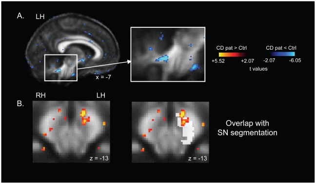
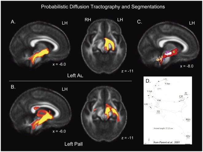
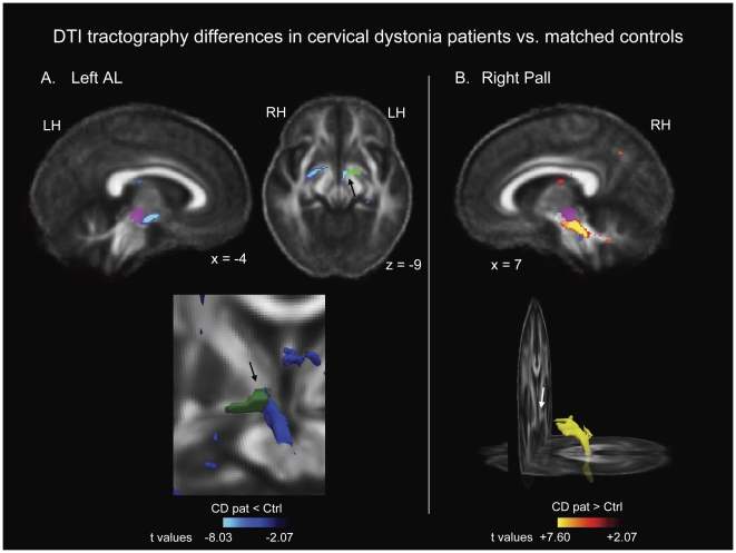
Methodology/principal findings: In the current study we investigated probabilistic diffusion tractography in DYT1-negative patients with cervical dystonia and matched healthy control subjects, with the goal of showing that patients exhibit altered microstructure in the connectivity between the pallidum and brainstem. The brainstem regions investigated included nuclei that are known to exhibit strong connections with the cerebellum. We observed large clusters of tractography differences in patients relative to healthy controls, between the pallidum and the brainstem. Tractography was decreased in the left hemisphere and increased in the right hemisphere in patients, suggesting a potential basis for the left/right white matter asymmetry we previously observed in focal dystonia patients.

Conclusions/significance: These findings support the hypothesis that connections between the basal ganglia and brainstem play a role in the pathophysiology of dystonia.


Conflict of interest statement

Competing Interests: The authors have declared that no competing interests exist.

Figures

Figure 1
Figure 1. Significant FA differences in cervical dystonia patients relative to control subjects.
(A) Reduced FA in the left cerebellar white matter in patients. (B) Increased FA adjacent to and overlapping with the left substantia nigra in patients. MNI talairach coordinates are indicated for each image. t maps are superimposed on the average FA map for all 24 subjects in the study, and are thresholded at t = +/−2.5 here for illustrative purposes. The color bar indicates the range of t values in this figure for FA contrasts, from +/−2.5 to the peak positive and negative t values for this contrast. Warm tones (red, orange, yellow) indicate regions in which cervical dystonia patients exhibited elevated FA relative to control subjects. Cool tones (blues) indicate regions in which cervical dystonia patients exhibited reduced FA relative to control subjects. LH: left hemisphere; RH: right hemisphere.
Figure 2
Figure 2. Probabilistic diffusion tractography and overlap with a priori areas of evaluation (AOEs) used for the FA and MD contrasts.
(A) Examples of tractography from the left ansa lenticularis (AL) seed region, averaged across control subjects (before contrasts were conducted), thresholded at 500 (samples). Note that tractography bifurcated at the level of the substantia nigra (see axial image). (B) Examples of tractography from the left pallidal seed region, averaged across control subjects (before contrasts were conducted), thresholded at 500. Like AL tractography, pallidal tractography bifurcated at the level of the substantia nigra (see axial image). There were also two projections to the thalamus, one superior and one more ventral (see sagittal image). (C) Intersection of tractography with segmentations of our a priori AOEs, including (in descending order) the ansa lenticularis (green), the substantia nigra (white), the red nucleus (pink), the pedunculopontine nucleus (blue), and the superior cerebellar peduncle (peach). Tractography maps are superimposed on the average FA map for all 24 subjects in the study. (D) Example of type Ib GPi neuron from , showing extensive arborization, including to the RN and PPN (reprinted with permission from The Journal of Comparative Neurology). MNI talairach coordinates are indicated for all images. LH: left hemisphere; RH: right hemisphere.
Figure 3
Figure 3. Regions of probabilistic diffusion tractography that were significantly different between cervical dystonia patients and controls.
(A) shows reduced tractography from the left AL seed region, and (B) shows elevated tractography from the right pallidal seed region. Arrows point to the region through which we drew ROIs in our previous DTI study, which includes AL fibers. A priori segmented regions are shown as reference points: Pink = red nucleus; White = substantia nigra; Blue = pedunculopontine nucleus. MNI talairach coordinates are indicated for each two dimensional image. Lower images in each panel show three dimensional rendering of clusters; the image for the left AL includes the AL seed region in green, and the patient/control difference is shown in blue. t maps and three dimensional clusters are superimposed on the average FA map for all 24 subjects in the study for anatomical reference, and are thresholded at t = +/−2.07 (the threshold used to identify difference clusters, p<0.05, uncorrected, for df = 22). The color bars indicate the range of t values in each panel, from +/−2.07 to the peak t value for each contrast. Warm tones (red, orange, yellow) indicate regions in which cervical dystonia patients exhibited elevated tractography relative to control subjects. Cool tones (blues) indicate regions in which cervical dystonia patients exhibited reduced tractography relative to control subjects. Three dimensional images are shown in mono-color rather than graded/multi-color to illustrate location rather than significance. LH: left hemisphere; RH: right hemisphere.

Similar articles

Cited by

References

    1. Loher TJ, Krauss JK. Dystonia associated with pontomesencephalic lesions. Mov Disord. 2009;24:157–167. - PubMed
    1. Tan EK, Chan LL, Auchus AP. Hemidystonia precipitated by acute pontine infarct. J Neurol Sci. 2005;234:109–111. - PubMed
    1. McNaught KS, Kapustin A, Jackson T, Jengelley TA, Jnobaptiste R, et al. Brainstem pathology in DYT1 primary torsion dystonia. Ann Neurol. 2004;56:540–547. - PubMed
    1. Wu CL, Lu CS. Delayed-onset dystonia following recovery from central pontine myelinolysis. J Formos Med Assoc. 1992;91:1013–1016. - PubMed
    1. Carbon M, Argyelan M, Habeck C, Ghilardi MF, Fitzpatrick T, et al. Increased sensorimotor network activity in DYT1 dystonia: a functional imaging study. Brain. 2010;133:690–700. - PMC - PubMed

Publication types