. 2012;7(2):e31677.
doi: 10.1371/journal.pone.0031677. Epub 2012 Feb 27.

Investigating the structural impacts of I64T and P311S mutations in APE1-DNA complex: a molecular dynamics approach

Affiliations

Investigating the structural impacts of I64T and P311S mutations in APE1-DNA complex: a molecular dynamics approach

C George Priya Doss et al. PLoS One. 2012.

Abstract

Background: Elucidating the molecular dynamic behavior of Protein-DNA complex upon mutation is crucial in current genomics. Molecular dynamics approach reveals the changes on incorporation of variants that dictate the structure and function of Protein-DNA complexes. Deleterious mutations in APE1 protein modify the physicochemical property of amino acids that affect the protein stability and dynamic behavior. Further, these mutations disrupt the binding sites and prohibit the protein to form complexes with its interacting DNA.

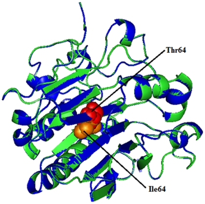
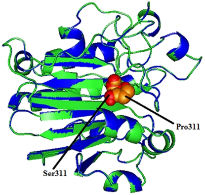
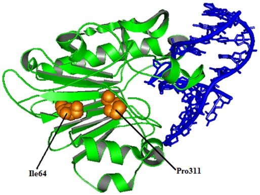
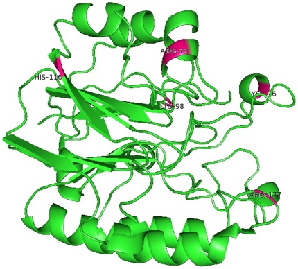
Principal findings: In this study, we developed a rapid and cost-effective method to analyze variants in APE1 gene that are associated with disease susceptibility and evaluated their impacts on APE1-DNA complex dynamic behavior. Initially, two different in silico approaches were used to identify deleterious variants in APE1 gene. Deleterious scores that overlap in these approaches were taken in concern and based on it, two nsSNPs with IDs rs61730854 (I64T) and rs1803120 (P311S) were taken further for structural analysis.

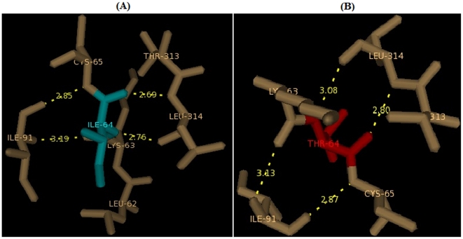
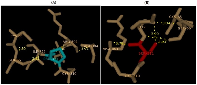
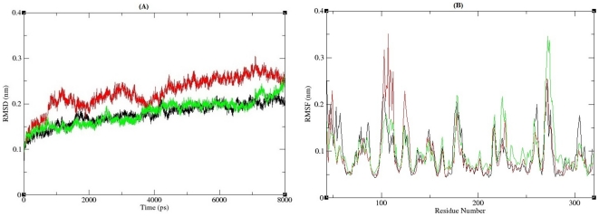
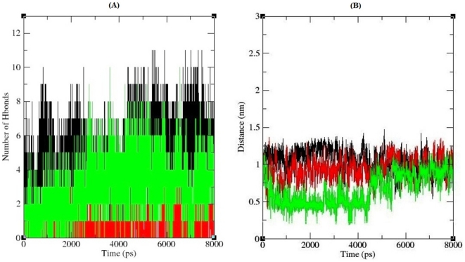
Significance: Different parameters such as RMSD, RMSF, salt bridge, H-bonds and SASA applied in Molecular dynamic study reveals that predicted deleterious variants I64T and P311S alters the structure as well as affect the stability of APE1-DNA interacting functions. This study addresses such new methods for validating functional polymorphisms of human APE1 which is critically involved in causing deficit in repair capacity, which in turn leads to genetic instability and carcinogenesis.


Conflict of interest statement

Competing Interests: The authors have declared that no competing interests exist.

Figures

Figure 1
Figure 1. Superimposed structure of native amino acid isoleucine in sphere shape (orange color) with mutant amino acid threonine (red color) at position 64 in PDB ID 1DE8 of APE1gene.
Figure 2
Figure 2. Superimposed structure of native amino acid proline in sphere shape (orange color) with mutant amino acid serine (red color) at position 311 in PDB ID 1DE8 of APE1 gene.
Figure 3
Figure 3. Changes in local environment and polar residue distance of APE1 protein brought about by an I64T mutation.
(A) The native type isoleucine residue (cyan) shows distance with surrounding residues.(B) Substitution of I64 residue to threnioine (red) shows surrounding residues and polar distance changes.
Figure 4
Figure 4. Changes in local environment and polar residue distance of APE1 protein brought about by P311S mutation.
(A) The native type proline residue (cyan) shows distance with surrounding residues. (B) Substitution of P311 residue to serine (red) shows surrounding residues and polar distance changes.
Figure 5
Figure 5. Co-crystal structure of APE1 protein with DNA and native amino acid isoleucine and proline at position 64 and 311 respectively.
Figure 6
Figure 6. RMSD and RMSF analysis of APE1-DNA complexes.
(A) Backbone RMSD of the APE1-DNA complexes. The ordinate is RMSD (nm), and the abscissa is time (ps). Black, Red and Green lines indicate native, I64T and P311S structures respectively. (B) RMSF of the carbon alpha atoms over the entire simulation. The ordinate is RMSF (nm), and the abscissa is residues. Black, Red and Green lines indicate native, I64T and P311S structures respectively.
Figure 7
Figure 7. Inter molecular hydrogen bond and salt bridge formation of APE1-DNA complexes.
(A) Number of hydrogen bond formed between APE1 protein and its binding DNA. Black, Red and Green lines indicate hydrogen bonds of the native, I64T and P311S structures respectively. (B) Stabilizing salt bridges formed between APE1 protein and DNA. The ordinate is distance (nm) and the abscissa is time (ps). Black, Red and Green lines indicate native, I64T and P311S complexes respectively.
Figure 8
Figure 8. Functional residues of APE1 protein involved in salt bridge formation (ARG 73, LYS 98, HIS 116, ARG 177 and LYS 276) were labeled and colored as pink.
Figure 9
Figure 9. Solvent accessible surface area (SASA) of APE1 protein.
Black, Red and Green lines indicate SASA of the native, I64T and P311S structures respectively. The ordinate is area (nm square) and the abscissa is time (ps).

Similar articles

Cited by

References

    1. Chang JS, Wrensch MR, Hansen HM, Sison JD, Aldrich MC, et al. Base excision repair genes and risk of lung cancer among San Francisco Bay Area Latinos and African-Americans. Carcinogenesis. 2009;30:78–87. - PMC - PubMed
    1. Bennett RA, Wilson DM, III, Wong D, Demple B. Interaction of human apurinic endonuclease and DNA polymerase beta in the base excision repair pathway. Proc Natl Acad Sci USA. 1997;94:7166–9. - PMC - PubMed
    1. Mol CD, Izumi T, Mitra S, Tainer JA. DNA-bound structures and mutants reveal abasic DNA binding by APE1 and DNA repair coordination. Nature. 2000;403:451–456. - PubMed
    1. Wilson DM, III, Bohr VA. The Mechanics of base excision repair and its relationship to aging and disease. DNA Repair (Amst.) 2007;6:544–559. - PubMed
    1. Yu E, Gaucher SP, Hadi MZ. Probing conformational changes in Ape1 during the progression of base excision repair. Biochemistry. 2010;49:3786–96. - PubMed