. 2012;7(2):e31742.
doi: 10.1371/journal.pone.0031742. Epub 2012 Feb 24.

Structural diversity in bacterial ribosomes: mycobacterial 70S ribosome structure reveals novel features

Affiliations

Structural diversity in bacterial ribosomes: mycobacterial 70S ribosome structure reveals novel features

Manidip Shasmal et al. PLoS One. 2012.

Abstract

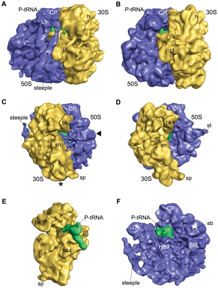
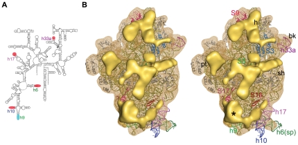
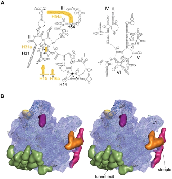
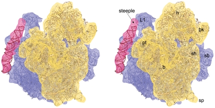
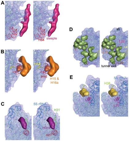
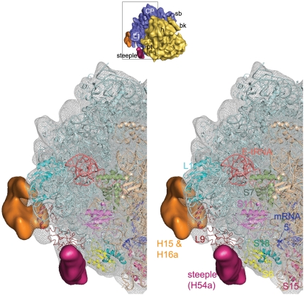
Here we present analysis of a 3D cryo-EM map of the 70S ribosome from Mycobacterium smegmatis, a saprophytic cousin of the etiological agent of tuberculosis in humans, Mycobacterium tuberculosis. In comparison with the 3D structures of other prokaryotic ribosomes, the density map of the M. smegmatis 70S ribosome reveals unique structural features and their relative orientations in the ribosome. Dramatic changes in the periphery due to additional rRNA segments and extra domains of some of the peripheral ribosomal proteins like S3, S5, S16, L17, L25, are evident. One of the most notable features appears in the large subunit near L1 stalk as a long helical structure next to helix 54 of the 23S rRNA. The sharp upper end of this structure is located in the vicinity of the mRNA exit channel. Although the M. smegmatis 70S ribosome possesses conserved core structure of bacterial ribosome, the new structural features, unveiled in this study, demonstrates diversity in the 3D architecture of bacterial ribosomes. We postulate that the prominent helical structure related to the 23S rRNA actively participates in the mechanisms of translation in mycobacteria.


Conflict of interest statement

Competing Interests: The authors have declared that no competing interests exist.

Figures

Figure 1
Figure 1. Comparison of the 70S Ribosome from M. smegmatis with the E. coli 70S ribosome.
The cryo-EM map of the Msm70S (A, C) is shown together with the cryo-EM map of the Eco70S (B, D; EMD-1395). The ribosomes are shown from the L1 side (A, and B) and the L7/L12 side (C,and D). Missing extended L7/L12 Stalk is marked in Msm70S with solid triangle (◂). Asterisk (*) marks the missing density in the bottom part of the 30S subunit of Msm70S due to shorter h10 and h17. The location of the S1 and S2 proteins where corresponding densities are largely absent in Msm70S is marked with arrow. The computationally separated small subunit (E) and large subunit (F) of the Msm70S are shown with the P site-bound tRNA (green) from the interface sides. The small subunits are shown in yellow, the large subunits blue in all the panels. Landmarks for the 30S subunit: bk, beak; h, head; pt, platform; sh, shoulder; sp, spur; h44, helix 44 of 16S rRNA. Landmarks for the 50S subunit: CP, central protuberance; L1, L1 protein; st, L7/L12 stalk; sb, L7/L12 stalk base; SRL, sarcin ricin loop; H69, helix 69 of 23S rRNA.
Figure 2
Figure 2. Structural analysis of the Msm30S.
(A) Secondary structure diagram of the M. tuberculocis 16S rRNA. The helices which are different in mycobacterial 16S rRNA as compared to the E. coli 16S rRNA are marked (shorter, red; longer, cyan). (B) Stereo view of the solvent side of Msm30S (yellow wire mesh) with the docked crystal structure of E. coli 30S subunit (16S rRNA in olive, proteins in grey colour) (pdb code: 2I2U). Major extra density clusters (solid yellow) are shown. Proteins with additional segments are coloured and designated with their names. Density cluster marked with asterisk (*) represents the density corresponding to extra components of h9 and proteins S16, S17. Landmarks are as in Figure 1.
Figure 3
Figure 3. Structural analysis of the Msm50S.
(A) Secondary structure diagram of the M. laprae 23S rRNA (left: 5′ end; right: 3′ end). Locations of extra rRNA helices in mycobacterium are highlighted (orange) and marked in the 5′ 23S rRNA structure (domains I, II, III). (B) Stereo view of the solvent side of Msm50S (blue wire mesh) with the coordinates of E. coli 50S subunit (23S rRNA pale cyan, 5S rRNA deep blue, proteins grey) docked inside. The atomic structure is adopted from the crystal structure of E. coli 70S ribosome (Protein Data Bank ID code 2I2V). Major additional density clusters are highlighted in different colors; steeple, deep pink; H15/H16a, orange; H31a, purple; additional density of L25, yellow; additional densities of proteins around the tunnel exit, green. Landmarks are as in Figure 1.
Figure 4
Figure 4. Location of the steeple (H54a).
Stereo representation of the density map of steeple is shown in wire mesh (deep pink) with 50S (blue) and 30S subunit (yellow) to show its orientation relative to the Msm70S subunits. Atomic coordinates of the E. coli 30S (pdb code: 2I2U) and 50S (pdb code: 2I2V) subunits docked into the corresponding density maps are also shown . A 96 nucleotide long rRNA helix has also been fitted into the density of steeple. Landmarks for the 30S subunit: bk, beak; h, head; pt, platform; sh, shoulder; sp, spur; b, body. Landmarks for the 50S subunit: L1, L1 protein; sb, L7/L12 stalk base.
Figure 5
Figure 5. Identification of the extra rRNA helices and additional segments of r-proteins in the Msm50S.
Close-up views of the (A) H54 region showing that the steeple (deep pink) emerges from H54, (B) H14 and H16 region displaying that the bifurcated density (orange) is related to these helices, and (C) H31 region identifying the density corresponding to the extra helix (purple), H31a, in the 23S rRNA, are shown in stereo. Density clusters attributable to extra domains of M. smegmatis ribosomal large subunit proteins L4/L17/L22/L29 (D), and L25 (E) are also shown in stereo. T. thermophilus L25 (from pdb code 2J01, chain Z, 3–179 amino acids) protein structure is shown here (magenta) to identify the C-terminal domain. Part of the C-terminal extra domain overlaps with the density cluster attributed to L25 in Msm70S.
Figure 6
Figure 6. Neighbourhood of the steeple structure.
Close-up view of the steeple (deep pink) is shown with its neighbouring ribosomal proteins and rRNA structures in stereo. The Msm70S density map is represented in grey wire mesh with the coordinates of the 30S (Wheat colour) and the 50S (light cyan) subunits docked into the map. Coordinate of mRNA (blue stick model) is taken from a crystal structure of the T. thermophilus 70S ribosome (pdb code: 2HGR, chain 1) and aligned to the atomic structures used here. A thumbnail view of the Msm70S is shown on top to orient the reader.

Similar articles

Cited by

References

    1. Schmeing TM, Ramakrishnan V. What recent ribosome structures have revealed about the mechanism of translation. Nature. 2009;461:1234–1242. - PubMed
    1. Baker R, Ridell M, Lind A, Ouchterlony O. Immunodiffusion studies of various structural preparations from mycobacterial cells. Int Arch Allergy Appl Immunol. 1979;59:328–336. - PubMed
    1. Baker RE, Hill WE, Larson CL. Delayed hypersensitivity reactions provoked by ribosomes from acid-fast bacilli. I. Ribosomal isolation, characterization, delayed hypersensitivity, and specificity. Infect Immun. 1972;6:258–265. - PMC - PubMed
    1. Baker RE, Hill WE, Larson CL. Ribosomes of acid-fast bacilli: immunogenicity, serology, and in vitro correlates of delayed hypersensitivity. Infect Immun. 1973;8:236–244. - PMC - PubMed
    1. Lonnroth I, Ridell M. Analyses of ribosomal proteins from mycobacteria with particular reference to the beta antigen. FEMS Microbiology Letters. 1985;27:203–207.

Publication types

MeSH terms