. 2012;7(2):e31856.
doi: 10.1371/journal.pone.0031856. Epub 2012 Feb 27.

A phylogenetic analysis of the globins in fungi

Affiliations

A phylogenetic analysis of the globins in fungi

David Hoogewijs et al. PLoS One. 2012.

Abstract

Background: All globins belong to one of three families: the F (flavohemoglobin) and S (sensor) families that exhibit the canonical 3/3 α-helical fold, and the T (truncated 3/3 fold) globins characterized by a shortened 2/2 α-helical fold. All eukaryote 3/3 hemoglobins are related to the bacterial single domain F globins. It is known that Fungi contain flavohemoglobins and single domain S globins. Our aims are to provide a census of fungal globins and to examine their relationships to bacterial globins.

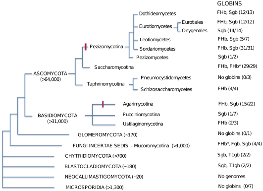
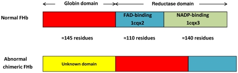
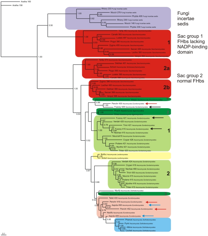
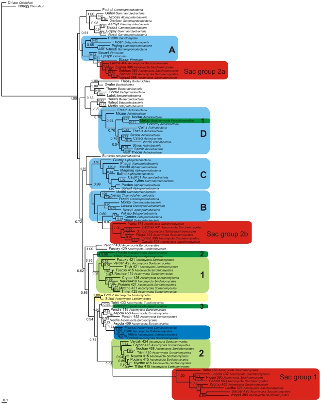
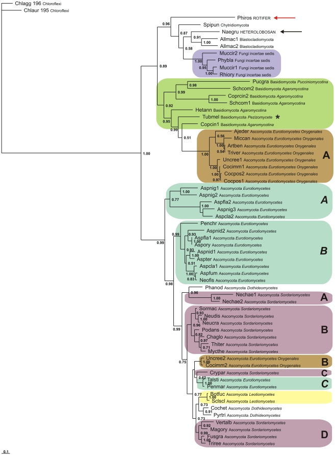
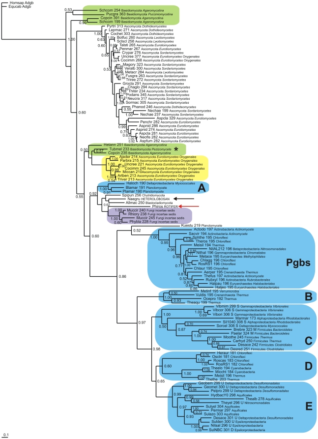
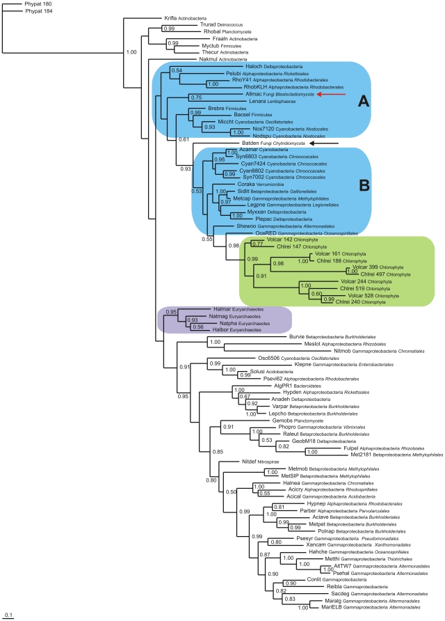
Results: Examination of 165 genomes revealed that globins are present in >90% of Ascomycota and ~60% of Basidiomycota genomes. The S globins occur in Blastocladiomycota and Chytridiomycota in addition to the phyla that have FHbs. Unexpectedly, group 1 T globins were found in one Blastocladiomycota and one Chytridiomycota genome. Phylogenetic analyses were carried out on the fungal globins, alone and aligned with representative bacterial globins. The Saccharomycetes and Sordariomycetes with two FHbs form two widely divergent clusters separated by the remaining fungal sequences. One of the Saccharomycete groups represents a new subfamily of FHbs, comprising a previously unknown N-terminal and a FHb missing the C-terminal moiety of its reductase domain. The two Saccharomycete groups also form two clusters in the presence of bacterial FHbs; the surrounding bacterial sequences are dominated by Proteobacteria and Bacilli (Firmicutes). The remaining fungal FHbs cluster with Proteobacteria and Actinobacteria. The Sgbs cluster separately from their bacterial counterparts, except for the intercalation of two Planctomycetes and a Proteobacterium between the Fungi incertae sedis and the Blastocladiomycota and Chytridiomycota.

Conclusion: Our results are compatible with a model of globin evolution put forward earlier, which proposed that eukaryote F, S and T globins originated via horizontal gene transfer of their bacterial counterparts to the eukaryote ancestor, resulting from the endosymbiotic events responsible for the origin of mitochondria and chloroplasts.


Conflict of interest statement

Competing Interests: The authors have declared that no competing interests exist.

Figures

Figure 1
Figure 1. Diagrammatic representation of fungal phylogeny based on ref. 18 and of their putative globins.
Estimates of the numbers of species in parentheses are from the Dictionary of the Fungi . Red bar indicates multicellularity with differentiated tissues; FHb* - unknown N-terminal domain linked to a FHb with an incomplete reductase domain, T1gb - T globin group1. Ratios refer to the number of genomes containing globins versus the total number of genomes analyzed.
Figure 2
Figure 2. Diagrammatic representation of FHb structures.
Schematically displayed structures are those of a normal FHb and of an abnormal chimeric FHb missing the C-terminal moiety of the reductase domain found in 21 of 29 Saccharomycotina genomes. The normal FHb is represented by E. coli FHb (PDB: 1gvh).
Figure 3
Figure 3. Bayesian phylogenetic tree of fungal FHbs.
Bayesian tree based on a MAFFT v.6.850 alignment of 62 fungal FHb globin domains using two plant nonsymbiotic Hbs as outgroup. Support values at branches represent Bayesian posterior probabilities (>0.5). The sequences are identified by the first three letters of the binary species name, the number of residues, and the full phylum and family names (see Table S1). Sac – Saccharomycetes.
Figure 4
Figure 4. Bayesian phylogenetic tree of fungal and bacterial FHbs.
Bayesian tree based on a T-COFFEE 9.01 alignment of the globin domains of 55 representative fungal FHbs and 54 representative bacterial FHbs. Support values at branches represent Bayesian posterior probabilities (>0.5). All the bacterial FHbs are in the blue boxes. The sequences are identified by the first three letters of the binary species name, the number of residues, and the full phylum and family names (see Table S1). Sac – Saccharomycetes.
Figure 5
Figure 5. Bayesian phylogenetic tree of fungal Sgbs.
Bayesian tree based on a MAFFT v.6.850 alignment of 59 fungal, one rotifer and one heterolobosan Sgbs, using two bacterial Pgbs as outgroup. Support values at branches represent Bayesian posterior probabilities (>0.5). The sequences are identified by the first three letters of the binary species name, and the full phylum and family names (see Table S1).
Figure 6
Figure 6. Bayesian phylogenetic tree of fungal and bacterial Sgbs.
Bayesian tree based on a T-COFFEE 9.01 alignment of 51 fungal, 57 bacterial (including 16 Pgbs), one rotifer and one heterolobosan Sgbs, using two Adgb sequences as outgroup. Support values at branches represent Bayesian posterior probabilities (>0.5). The sequences are identified by the first three letters of the binary species name, the number of residues, and the full phylum and family names (see Table S1).
Figure 7
Figure 7. Bayesian phylogenetic tree of fungal and bacterial T1 globins.
Bayesian tree based on a T-COFFEE 9.01 alignment of 2 fungal (red and black arrows), 70 bacterial (blue), 4 euryarchaeote (purple) and 10 chlorophyte (green) T1 globins, using 2 Physcomitrella nsHbs as outgroup. Support values at branches represent Bayesian posterior probabilities (>0.5). All the bacterial FHbs are in the blue boxes. The sequences are identified by the first three letters of the binary species name, the number of residues, and the full phylum and family names (see Table S1).

Similar articles

  • Bacterial and archaeal globins - a revised perspective.
    Vinogradov SN, Tinajero-Trejo M, Poole RK, Hoogewijs D. Vinogradov SN, et al. Biochim Biophys Acta. 2013 Sep;1834(9):1789-800. doi: 10.1016/j.bbapap.2013.03.021. Epub 2013 Mar 27. Biochim Biophys Acta. 2013. PMID: 23541529
  • Three globin lineages belonging to two structural classes in genomes from the three kingdoms of life.
    Vinogradov SN, Hoogewijs D, Bailly X, Arredondo-Peter R, Guertin M, Gough J, Dewilde S, Moens L, Vanfleteren JR. Vinogradov SN, et al. Proc Natl Acad Sci U S A. 2005 Aug 9;102(32):11385-9. doi: 10.1073/pnas.0502103102. Epub 2005 Aug 1. Proc Natl Acad Sci U S A. 2005. PMID: 16061809 Free PMC article.
  • A phylogenomic profile of globins.
    Vinogradov SN, Hoogewijs D, Bailly X, Arredondo-Peter R, Gough J, Dewilde S, Moens L, Vanfleteren JR. Vinogradov SN, et al. BMC Evol Biol. 2006 Apr 7;6:31. doi: 10.1186/1471-2148-6-31. BMC Evol Biol. 2006. PMID: 16600051 Free PMC article.
  • Microbial eukaryote globins.
    Vinogradov SN, Bailly X, Smith DR, Tinajero-Trejo M, Poole RK, Hoogewijs D. Vinogradov SN, et al. Adv Microb Physiol. 2013;63:391-446. doi: 10.1016/B978-0-12-407693-8.00009-1. Adv Microb Physiol. 2013. PMID: 24054801 Review.
  • Globin phylogeny, evolution and function, the newest update.
    Schuster CD, Salvatore F, Moens L, Martí MA. Schuster CD, et al. Proteins. 2024 Jun;92(6):720-734. doi: 10.1002/prot.26659. Epub 2024 Jan 9. Proteins. 2024. PMID: 38192262 Review.

Cited by

References

    1. Keilin D. Occurrence of haemoglobin in yeast and the supposed stabilization of the oxygenated cytochrome oxidase. Nature. 1953;172:390–393. - PubMed
    1. Keilin D, Tissieres A. Haemoglobin in moulds Neuropora crassa and Penicillium notatum. Nature. 1953;172:393–394. - PubMed
    1. Oshino R, Asakura T, Takio K, Oshino N, Chance B, et al. Purification and molecular properties of yeast hemoglobin. Eur J Biochem. 1973;39:581–590. - PubMed
    1. Iwaasa H, Takagi T, Shikama K. Amino acid sequence of yeast hemoglobin. A twodomain structure. J Mol Biol. 1992;227:948–954. - PubMed
    1. Zhu H, Riggs A. Yeast flavohemoglobin is an ancient protein related to globins and a reductase family. Proc Natl Acad Sci USA. 1992;89:5015–5019. - PMC - PubMed

Publication types