Evaluation of functional erythropoietin receptor status in skeletal muscle in vivo: acute and prolonged studies in healthy human subjects
- PMID: 22384088
- PMCID: PMC3285196
- DOI: 10.1371/journal.pone.0031857
Evaluation of functional erythropoietin receptor status in skeletal muscle in vivo: acute and prolonged studies in healthy human subjects
Abstract
Background: Erythropoietin receptors have been identified in human skeletal muscle tissue, but downstream signal transduction has not been investigated. We therefore studied in vivo effects of systemic erythropoietin exposure in human skeletal muscle.
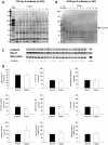
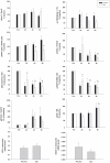
Methodology/principal findings: The protocols involved 1) acute effects of a single bolus injection of erythropoietin followed by consecutive muscle biopsies for 1-10 hours, and 2) a separate study with prolonged administration for 16 days with biopsies obtained before and after. The presence of erythropoietin receptors in muscle tissue as well as activation of Epo signalling pathways (STAT5, MAPK, Akt, IKK) were analysed by western blotting. Changes in muscle protein profiles after prolonged erythropoietin treatment were evaluated by 2D gel-electrophoresis and mass spectrometry. The presence of the erythropoietin receptor in skeletal muscle was confirmed, by the M20 but not the C20 antibody. However, no significant changes in phosphorylation of the Epo-R, STAT5, MAPK, Akt, Lyn, IKK, and p70S6K after erythropoietin administration were detected. The level of 8 protein spots were significantly altered after 16 days of rHuEpo treatment; one isoform of myosin light chain 3 and one of desmin/actin were decreased, while three isoforms of creatine kinase and two of glyceraldehyd-3-phosphate dehydrogenase were increased.
Conclusions/significance: Acute exposure to recombinant human erythropoietin is not associated by detectable activation of the Epo-R or downstream signalling targets in human skeletal muscle in the resting situation, whereas more prolonged exposure induces significant changes in the skeletal muscle proteome. The absence of functional Epo receptor activity in human skeletal muscle indicates that the long-term effects are indirect and probably related to an increased oxidative capacity in this tissue.
Conflict of interest statement
Figures


Similar articles
-
Erythropoietin receptor in human skeletal muscle and the effects of acute and long-term injections with recombinant human erythropoietin on the skeletal muscle.J Appl Physiol (1985). 2008 Apr;104(4):1154-60. doi: 10.1152/japplphysiol.01211.2007. Epub 2008 Jan 24. J Appl Physiol (1985). 2008. PMID: 18218911
-
Erythropoietin does not activate erythropoietin receptor signaling or lipolytic pathways in human subcutaneous white adipose tissue in vivo.Lipids Health Dis. 2016 Sep 17;15(1):160. doi: 10.1186/s12944-016-0327-z. Lipids Health Dis. 2016. PMID: 27640183 Free PMC article.
-
Prolonged erythropoietin treatment does not impact gene expression in human skeletal muscle.Muscle Nerve. 2015 Apr;51(4):554-61. doi: 10.1002/mus.24355. Epub 2015 Feb 25. Muscle Nerve. 2015. PMID: 25088500
-
STAT5 as a Key Protein of Erythropoietin Signalization.Int J Mol Sci. 2021 Jul 1;22(13):7109. doi: 10.3390/ijms22137109. Int J Mol Sci. 2021. PMID: 34281163 Free PMC article. Review.
-
Erythropoietin: physiology and pharmacology update.Exp Biol Med (Maywood). 2003 Jan;228(1):1-14. doi: 10.1177/153537020322800101. Exp Biol Med (Maywood). 2003. PMID: 12524467 Review.
Cited by
-
Erythropoietin Reduces Insulin Resistance via Regulation of Its Receptor-Mediated Signaling Pathways in db/db Mice Skeletal Muscle.Int J Biol Sci. 2017 Oct 17;13(10):1329-1340. doi: 10.7150/ijbs.19752. eCollection 2017. Int J Biol Sci. 2017. PMID: 29104499 Free PMC article.
-
[Anatomical heterogeneity in the proteome of human subcutaneous adipose tissue].An Pediatr (Barc). 2013 Mar;78(3):140-8. doi: 10.1016/j.anpedi.2012.10.010. Epub 2012 Nov 24. An Pediatr (Barc). 2013. PMID: 23228439 Free PMC article. Spanish.
-
Erythropoietin Does Not Enhance Skeletal Muscle Protein Synthesis Following Exercise in Young and Older Adults.Front Physiol. 2016 Jul 8;7:292. doi: 10.3389/fphys.2016.00292. eCollection 2016. Front Physiol. 2016. PMID: 27458387 Free PMC article.
-
Erythropoietin doping in cycling: lack of evidence for efficacy and a negative risk-benefit.Br J Clin Pharmacol. 2013 Jun;75(6):1406-21. doi: 10.1111/bcp.12034. Br J Clin Pharmacol. 2013. PMID: 23216370 Free PMC article.
-
The role and regulation of erythropoietin (EPO) and its receptor in skeletal muscle: how much do we really know?Front Physiol. 2013 Jul 15;4:176. doi: 10.3389/fphys.2013.00176. eCollection 2013. Front Physiol. 2013. PMID: 23874302 Free PMC article.
References
-
- Fisher JW. Erythropoietin: physiology and pharmacology update. Exp Biol Med (Maywood) 2003;228:1–14. - PubMed
-
- Chong ZZ, Kang JQ, Maiese K. Erythropoietin: cytoprotection in vascular and neuronal cells. Curr Drug Targets Cardiovasc Haematol Disord. 2003;3:141–154. - PubMed
-
- Constantinescu SN, Ghaffari S, Lodish HF. The Erythropoietin Receptor: Structure, Activation and Intracellular Signal Transduction. Trends Endocrinol Metab. 1999;10:18–23. - PubMed
-
- Tilbrook PA, Klinken SP. The erythropoietin receptor. Int J Biochem Cell Biol. 1999;31:1001–1005. - PubMed
-
- Jelkmann W. Molecular biology of erythropoietin. Intern Med. 2004;43:649–659. - PubMed
Publication types
MeSH terms
Substances
LinkOut - more resources
Full Text Sources
Research Materials
Miscellaneous

