. 2012;7(2):e31959.
doi: 10.1371/journal.pone.0031959. Epub 2012 Feb 22.

Cytotoxicity of CD56(bright) NK cells towards autologous activated CD4+ T cells is mediated through NKG2D, LFA-1 and TRAIL and dampened via CD94/NKG2A

Affiliations

Cytotoxicity of CD56(bright) NK cells towards autologous activated CD4+ T cells is mediated through NKG2D, LFA-1 and TRAIL and dampened via CD94/NKG2A

Natasja Nielsen et al. PLoS One. 2012.

Abstract

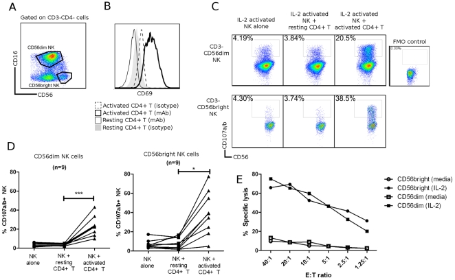
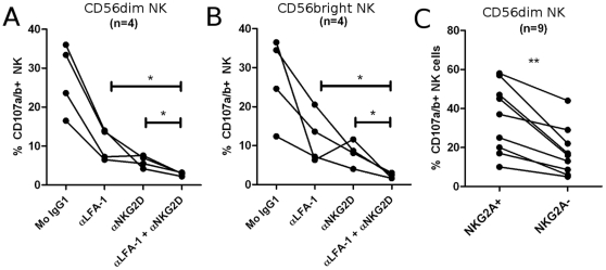
In mouse models of chronic inflammatory diseases, Natural Killer (NK) cells can play an immunoregulatory role by eliminating chronically activated leukocytes. Indirect evidence suggests that NK cells may also be immunoregulatory in humans. Two subsets of human NK cells can be phenotypically distinguished as CD16(+)CD56(dim) and CD16(dim/-)CD56(bright). An expansion in the CD56(bright) NK cell subset has been associated with clinical responses to therapy in various autoimmune diseases, suggesting an immunoregulatory role for this subset in vivo. Here we compared the regulation of activated human CD4(+) T cells by CD56(dim) and CD56(bright) autologous NK cells in vitro. Both subsets efficiently killed activated, but not resting, CD4(+) T cells. The activating receptor NKG2D, as well as the integrin LFA-1 and the TRAIL pathway, played important roles in this process. Degranulation by NK cells towards activated CD4(+) T cells was enhanced by IL-2, IL-15, IL-12+IL-18 and IFN-α. Interestingly, IL-7 and IL-21 stimulated degranulation by CD56(bright) NK cells but not by CD56(dim) NK cells. NK cell killing of activated CD4(+) T cells was suppressed by HLA-E on CD4(+) T cells, as blocking the interaction between HLA-E and the inhibitory CD94/NKG2A NK cell receptor enhanced NK cell degranulation. This study provides new insight into CD56(dim) and CD56(bright) NK cell-mediated elimination of activated autologous CD4(+) T cells, which potentially may provide an opportunity for therapeutic treatment of chronic inflammation.


Conflict of interest statement

Competing Interests: NN, BU, and PS are employees of Novo Nordisk A/S and own shares. This project was partly supported by Novo Nordisk A/S. PS has assigned patents related to NKG2A and NNC0141-0100 (WO2007042573, 20070726; WO2007147898, 20071227; WO2008009545, 20080124; WO2009092805, 20090730). BU has a patent related to NKG2D (WO09077483, 20090625). LLL and UCSF have licensed intellectual property rights regarding NKG2D for commercial applications. This does not alter the authors' adherence to all the PLoS ONE policies on sharing data and materials.

Figures

Figure 1
Figure 1. Activated NK cells kill activated, but not resting, CD4+ T cells.
(A) Representative gating strategy. NK cells were defined as viable, CD3 singlets. NK cell subsets were defined based on expression of CD16 and CD56. (B) Activation of CD4+ T cells was confirmed by CD69 upregulation. CD4+ T cells were activated for 4 days with anti-CD3+anti-CD28 Dynabeads (propionate was added on day 3). Resting CD4+ T cells were unstimulated in media for 4 days. (C–D) NK cells were cultured for 4 days in IL-2, and CD4+ T cells were activated as described. Resting CD4+ T cells were unstimulated for 4 days in culture. Autologous NK cells and CD4+ T cells were co-cultured at an E∶T ratio of 1∶1 for 4 hours with FITC-conjugated anti-CD107a+anti-CD107b mAb. Flow cytometry was performed to determine CD107a/b expression on NK cell subsets. Data shown in (C) are for a representative donor, (D) are for n = 9. Data represent mean ± SEM. * P<0.05; *** P<0.001. (E) Sorted CD56dim (□/▪) and CD56bright (○/•) NK cells were cultured in media (□/○) or IL-2 (▪/•) for 4 days, and co-cultured with 51Cr-labeled activated CD4+ T cells in a 51Cr-release assay. Experiment shown is representative of n = 3.
Figure 2
Figure 2. CD56bright NK cells have a higher cytotoxic potential towards activated CD4+ T cells.
Autologous NK cells and CD4+ T cells were isolated, and CD4+ T cells were activated for 4 days as described. NK cells were activated as indicated: 200 IU/mL IL-2, 25 ng/mL IL-4, 25 ng/mL IL-7, 25 ng/mL IL-9, 5 ug/mL IL-15, 100 ug/mL IL-21, 50 ug/mL IL-12, 0.25 mg/mL IL-18 or 100 U/mL IFN-αA. NK cells and CD4+ T cells were co-cultured for 4 hours with FITC-conjugated anti-CD107a+anti-CD107b antibodies. Flow cytometry was performed to determine degranulation of (A) CD56dim NK cells and (B) CD56bright NK cells. Data represent mean ± SEM of n≥4 experiments. Statistical significance is calculated in comparison to resting NK cells (media) co-cultured with activated CD4+ T cells. * P<0.05, ** P<0.005, *** P<0.001.
Figure 3
Figure 3. Multiple receptors and ligands are involved in NK cell-mediated lysis of activated CD4+ T cells.
Role of (A) activating and (B) inhibitory NK receptors in NK cell degranulation. Left column: representative histograms (of n≥3) for surface expression of ligands on activated (thick black line) and resting CD4+ T cells (thin black line). Isotype-matched control Ig are represented by dashed line (activated CD4+ T) and filled histogram (resting CD4+ T). Middle- and right column: NK and CD4+ T cells were activated for 4 days in vitro as described, and co-cultured for 4 hours with 10 ug/mL mAb (or relevant isotype-matched control Ig). Degranulation is shown for CD56dim (middle column) and CD56bright (right column) NK cells. Representative histograms of surface expression of receptors on activated (thick black line) and resting NK cells (thin black line). Isotype-matched control Ig are represented by dashed line (activated NK) and filled histogram (resting NK). * P<0.05, ** P<0.005, *** P<0.001. (C) Sorted IL-2-activated CD56dim and CD56bright NK cells were co-cultured with 51Cr-labeled activated CD4+ T cells in a 51Cr-release assay with human IgG4 isotype control (•) or anti-NKG2A mAb (○). Data represents n = 3 experiments.
Figure 4
Figure 4. NKG2D and LFA-1 act synergistically in mediating NK cell degranulation.
NK and CD4+ T cells were activated for 4 days in vitro as described, and co-cultured for 4 hours with FITC conjugated anti-CD107a+anti-CD107b mAbs. 10 ug/mL blocking antibodies (or relevant isotype-matched control Ig) were added where indicated. Flow cytometry was performed to assess degranulation of (A) CD56dim and (B) CD56bright NK cells. (C) CD56dim NK cells were gated based on surface expression of CD94/NKG2A, and degranulation of each subset was evaluated. * P<0.05.
Figure 5
Figure 5. NK cells mediate TRAIL-dependent cytotoxicity towards activated CD4+ T cells.
Autologous NK cells and CD4+ T cells were isolated and activated for 4 days as described. Resting NK and CD4+ T cells were unstimulated in media for 4 days. (A) Representative histograms for flow cytometric analysis of surface expression of TRAIL-R1 (DR4) and TRAIL-R2 (DR5) on activated CD4+ T cells (thick black line) and resting CD4+ T cells (thin black line). Isotype-matched control Ig are represented by dashed line (activated CD4+ T) and filled histogram (resting CD4+). (B) Histograms are representative of TRAIL surface expression on activated NK cells (thick black line) and resting NK cells (thin black line). Isotype-matched control Ig are represented by dashed line (activated NK) and filled histogram (resting NK). (C) Sorted IL-2-activated CD56dim and CD56bright NK cells were co-cultured with 51Cr-labeled activated CD4+ T cells in a 51Cr-release assay with isotype-matched control Ig (•) or anti-TRAIL mAb (○). Experiment shown is representative of n = 3.

References

    1. Trinchieri G. Biology of natural killer cells. Adv Immunol. 1989;47:187–376. - PMC - PubMed
    1. Vivier E, Tomasello E, Baratin M, Walzer T, Ugolini S. Functions of natural killer cells. Nat Immunol. 2008;9:503–510. - PubMed
    1. Lanier LL. NK cell recognition. Annu Rev Immunol. 2005;23:225–274. - PubMed
    1. Ljunggren HG, Karre K. In search of the ‘missing self’: MHC molecules and NK cell recognition. Immunol Today. 1990;11:237–244. - PubMed
    1. Zhang B, Yamamura T, Kondo T, Fujiwara M, Tabira T. Regulation of experimental autoimmune encephalomyelitis by natural killer (NK) cells. J Exp Med. 1997;186:1677–1687. - PMC - PubMed

Publication types

MeSH terms

Substances